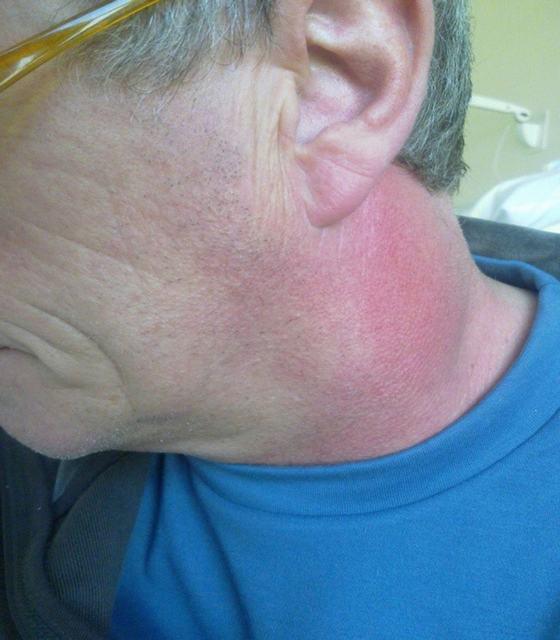
Chin abscess. Odontogenic phlegmon of the submandibular region on the right
Odontogenic abscesses and phlegmons of the maxillary fossa are common, as they can occur with any disease from the group of odontogenic infections - periodontitis, periostitis, osteomyelitis, with retention and dystopia of teeth, festering cysts, alveolitis, etc.
Abscess- This is a limited purulent inflammation of the soft tissues.
Phlegmon- diffuse purulent-necrotic inflammation of cellular spaces, subcutaneous fat, interfascial spaces and other soft tissues. Distinguish purulent, anaerobic or putrefactive phlegmon.
The causative agent of abscesses and phlegmon are staphylococci, streptococci, rarely Pseudomonas aeruginosa, Escherichia coli, anaerobes.
The spread of infection often occurs by contact, along the length or with the flow of lymph.
The onset of the disease is often preceded by an acute respiratory infection, flu, tonsillitis, hypothermia, overheating, stress, anemia, tooth extraction, trauma, etc.
In the clinic of abscesses and phlegmon, acute and subacute stages are distinguished. The acute stage is characterized by an increase in local signs of inflammation (edema, hyperemia, pain, formation of infiltrates, dysfunction), pronounced general reactions of the body in the form of fever, fever, malaise, headache, leukocytosis in the blood. If timely opening of the abscess does not occur (through a fistula or surgically), the infectious and inflammatory process can spread to neighboring anatomical regions, into the cranial cavity, into the deep cellular spaces of the neck, and the mediastinum. In this regard, complications such as thrombosis of the sinuses of the dura mater, meningoencephalitis, mediastinitis, secondary destructive osteomyelitis of the jaws, and sepsis may develop.
Treatment consists in a wide opening and drainage of the purulent focus, sometimes it is necessary to make several incisions in the maxillary fossa, daily washing of the purulent wound with antiseptic solutions, passive and active immunization, the introduction of desensitizing therapy and hormone therapy, and detoxification infusion therapy. Normalization of water-salt metabolism is carried out.
Every day it is necessary to monitor the condition of the wound and general well-being, blood pressure, temperature, diuresis, personal hygiene. When acute inflammatory manifestations subside, physiotherapeutic treatment is prescribed (electrophoresis, UHF, microwave, etc.).
The nutrition of such patients should be high-calorie, sparing, rich in vitamins.
Currently, there are several classification schemes for phlegmon of the maxillofacial area. From the point of view of practical dentistry, it is advisable to apply the Evdokimov scheme, built on topographic and anatomical principles:
- Abscesses and phlegmon, localized in the region of the upper jaw:
- infraorbital region;
- zygomatic area;
- orbital area;
- temporal fossa;
- infratemporal and pterygopalatine fossae.
- Abscesses and phlegmon, localized in the lower jaw:
- chin area;
- buccal area;
- submandibular region;
- peripharyngeal space;
- pterygoid-maxillary space;
- areas of the parotid salivary gland and the retromaxillary fossa.
- Abscesses and phlegmon of the floor of the mouth.
- Abscesses and phlegmon of the neck (superficial and deep).
Borders of the infraorbital region: upper - lower edge of the orbit, lower - alveolar process of the upper jaw; internal - the edge of the pear-shaped opening; external - zygomatic-maxillary suture.
foci of infection in the periodontium 543 | 345 teeth, wounds, infectious and inflammatory lesions of the skin of the infraorbital region, infection during infected anesthesia.
Symptoms: severe throbbing pain, swelling of the tissues of the infraorbital region, eyelids, infiltrate, which is determined in the region of the arch of the vestibule of the mouth, pain on palpation, fluctuation during the maturation of the abscess.
Abscesses and phlegmon of the zygomatic region
The boundaries of the zygomatic region: upper - anteroinferior part of the temporal region and the lower edge of the orbit; lower - anterior-upper section of the buccal region; anterior - zygomatic-maxillary suture; posterior - zygomatic-temporal suture.
The main sources and ways of infection: foci of infection in the periodontium 654 | 456 teeth, wounds, infectious and inflammatory processes of the skin of the zygomatic region, infection during infiltration anesthesia, spread of infection from the buccal and infraorbital region.
Symptoms: infiltration of the tissues of the zygomatic region, swelling of the eyelids, hyperemia of the skin, fluctuation during suppuration, moderate pain, limited mouth opening, moderate intoxication.
Abscesses and phlegmon of the orbit
Region boundaries: walls of the eyeball.
The main sources and ways of infection: foci of periodontal infection 543 | 345 teeth, wounds, infectious and inflammatory lesions of the skin and eyelids, the spread of infection along the length of the maxillary sinus, infraorbital region, zygomatic region, infratemporal and pterygopalatine fossae.
Symptoms: severe swelling of the eyelids and conjunctiva; exophthalmos, limited movement of the eyeball, diplopia, partial or complete blindness, general reaction in the form of leukocytosis, fever, symptoms of intoxication.
Abscesses and phlegmon of the buccal region
Region boundaries: upper - the lower edge of the zygomatic bone, lower - the lower edge of the lower jaw, anterior - the line connecting the zygomatic-maxillary suture with the angle of the mouth, posterior - the anterior edge of the masticatory muscle.
In this area, superficial and deep phlegmons and abscesses are distinguished (in relation to the buccal muscle).
The main sources of infection: foci of infection in the periodontium of the molars and premolars of both jaws, wounds, infectious and inflammatory processes along the length of the infraorbital, zygomatic and parotid-masticatory regions.
Symptoms: infiltration of the tissues of the buccal region and eyelids; hyperemia and skin tension over the infiltrate; pain, aggravated by palpation of the infiltrate and opening the mouth; fluctuation in the center of the infiltrate, the general condition is satisfactory, with deep phlegmon and abscesses, local signs of inflammation appear in the oral cavity.
Abscesses and phlegmon of the infratemporal region
Borders of the infratemporal fossa: upper - infratemporal crest of the main bone, lower - buccal-pharyngeal fascia, anterior - tubercle of the upper jaw and zygomatic bone, posterior - styloid process with muscles attached to it, outer - inner surface of the lower jaw branch.
The main sources and ways of infection: foci of infection in the periodontium 87 | 78 teeth, infection during conduction anesthesia at the tubercle of the upper jaw, spread of infection along the length of the pterygo-maxillary space, buccal region.
Symptoms: severe pain in the area of the infiltrate, even at rest, radiating to the corresponding half of the head, aggravated by opening the mouth; local signs of inflammation are not pronounced due to a deeply located infiltrate; swelling of soft tissues above and below the zygomatic arch; in the oral cavity, the infiltrate is located in the posterior sections of the arch of the vestibule of the mouth, painful on palpation; the mucosa over it is hyperemic; symptoms of intoxication are expressed.
Abscesses and phlegmon of the temporal region
The boundaries of the temporal region: the upper and back are the temporal line of the frontal and parietal bones, the lower is the infratemporal crest of the main bone, the inner is the temporal platform formed by the frontal, temporal, parietal and main bones, the outer is the zygomatic arch.
There are superficial abscesses and phlegmons located between the skin and the temporal aponeurosis, between the temporal aponeurosis and the temporal muscle, and deep ones, located between the temporal muscle and the bottom of the temporal bone.
The main sources and ways of infection: wounds and infectious and inflammatory lesions of the skin of the temporal region, the spread of infection from the infratemporal fossa, buccal region, parotid-masticatory region.
Symptoms: with superficial localization of the purulent focus, a pronounced swelling of the soft tissues of the temporal region, skin hyperemia, pain, aggravated by palpation, and fluctuation appear.
With deep phlegmon and abscesses, severe spontaneous pain, inflammatory contracture of the jaw, moderate swelling and hyperemia of the skin come to the fore, symptoms of intoxication are expressed.
Abscesses and phlegmon of the parotid-masticatory region
Borders: upper - the lower edge of the zygomatic bone of the zygomatic arch, lower - the lower edge of the body of the lower jaw, anterior - the anterior edge of the chewing region, posterior - the posterior edge of the lower jaw branch.
Superficial phlegmons and abscesses are located between the skin and the parotid-masticatory fascia and the outer surface of the lower jaw branch.
Deep abscesses and phlegmon are located between the chewing muscle and the outer surface of the lower jaw branch.
The main sources and ways of infection: foci of odontogenic infection in the area of the third molars, wounds, infectious and inflammatory processes of the skin of the parotid-masticatory region, the spread of infection from the buccal region, retromaxillary, submandibular, parotid salivary gland.
Symptoms: with superficial abscesses and phlegmon, a sharp swelling of the soft tissues of the parotid-masticatory region, hyperemia of the skin over the infiltrate, pain, aggravated by palpation and opening the mouth, fluctuation, moderate contracture of the jaw. With deep phlegmon and abscesses - intense pain when opening the mouth and at rest, severe jaw contracture, moderate soft tissue swelling, more pronounced general symptoms of inflammation.
Abscesses and phlegmon of the posterior maxillary region
Region boundaries: upper - external auditory canal, lower - lower pole of the parotid gland, anterior - posterior edge of the lower jaw branch, posterior - mastoid process of the temporal bone and sternocleidomastoid muscle, internal - styloid process of the temporal bone with muscles attached to it; external - parotid-chewing fascia.
The main sources and ways of infection: wounds and infectious and inflammatory lesions of the skin of the posterior maxillary region, the spread of infection from the parotid-masticatory region, submandibular, pterygo-maxillary space, parotid salivary gland.
Symptoms: pain in the mandibular region, aggravated by opening the mouth, swelling of the soft tissues, tension and hyperemia of the skin over the infiltrate, fluctuation, moderate contracture of the jaws, general signs of inflammation.
Abscesses and phlegmon of the pterygo-jaw space
Borders: the outer - the inner surface of the lower jaw branch and the lower - the temporal muscle, the inner, posterior and lower - the outer surface of the medial pterygoid muscle, the upper - the external pterygoid muscle, the anterior - the buccal-pharyngeal suture.
The main sources and ways of infection: foci of periodontal infection of the third molars of the lower jaw, infection during conduction anesthesia of the lower alveolar nerve, spread of infection from the palatine tonsils.
Symptoms: limited opening of the mouth, sore throat, aggravated by swallowing, deep palpation of the supramaxillary region can reveal infiltration, hyperemia and swelling of the oral mucosa in the area of the pterygomandibular fold, severe aseptic condition, intoxication.
Abscesses and phlegmon of the peripharyngeal space
Borders: external - medial pterygoid muscle, internal - lateral wall of the pharynx and muscle that lifts and stretches the soft palate, anterior - interpterygoid fascia, posterior - lateral facies spurs extending from the prevertebral fascia to the pharyngeal wall, lower - submandibular salivary gland.
 |
| Rice. a - frontal plane: 1 - chewing muscle; 2- medial pterygoid muscle; 3 - lateral pterygoid muscle; 4 - temporal muscle; 6 - lower jaw; 7 - lateral wall of the pharynx; b - horizontal plane: 1 - chewing muscle; 2 - medial pterygoid muscle; 3 - parotid gland; 4 - pharyngeal-prevertebral fascia; 5 - inflammatory infiltrate; 6 - lower jaw; 7 - stylodiaphragm; 8 - palatine tonsil; 9 - internal carotid artery; 10 - internal jugular vein; 11 - posterior peripharyngeal space |
The main sources and ways of infection: wounds, infectious and inflammatory processes of the pharyngeal mucosa, the spread of infection from the pterygo-maxillary space, submandibular region, sublingual, parotid-masticatory and retromaxillary regions, from the side of the palatine tonsils.
Symptoms: sore throat when swallowing and at rest, shortness of breath, pronounced swelling of the soft tissues of the submandibular region, the infiltrate is located deep, can be palpated in the angle of the lower jaw, painful, swelling of the side wall of the oropharynx, the pharynx is asymmetric, the general condition is severe, contracture of the lower jaw is expressed.
Borders: the upper one is the mucous membrane of the bottom of the mouth, the lower one is the maxillo-hyoid muscle, the outer one is the inner surface of the body of the lower jaw, the inner one is the genio-lingual and genio-hyoid muscle.
The main sources and ways of infection: foci of infection in the periodontal teeth of the lower jaw, more often in the region of premolars and molars, wounds and infectious and inflammatory lesions of the mucous membrane of the sublingual region, the excretory duct of the submandibular salivary gland.
Symptoms: pain in the sublingual region, aggravated by swallowing, talking, moving the tongue, palpation. The appearance of the patient is characteristic: the mouth is half open, saliva flows out, a putrid odor comes from the mouth. Mouth opening is limited. The tongue is covered with a dirty gray coating, raised. The mucous membrane of the bottom of the oral cavity is hyperemic, edematous. General condition of moderate severity.
Abscesses and phlegmon of the sublingual region
Phlegmon of the floor of the mouth. Phlegmon of the floor of the mouth is a diffuse purulent inflammation of the tissues located above and below the muscular diaphragm of the floor of the mouth (sublingual and submandibular regions).
The main sources and ways of infection: foci of infection in the periodontium of the teeth of the lower jaw, wounds, infectious and inflammatory lesions of the mucous membrane of the bottom of the cavity, the skin of the chin area and submandibular area, behind the jaw and peripharyngeal space.
Symptoms: pain aggravated by swallowing, talking, palpation of the infiltrate, shortness of breath, up to asphyxia, forced position of the patient (he sits with his head tilted forward, a suffering look, his mouth is half open, saliva flows out of it, speech is slurred, an unpleasant odor comes from his mouth); the infiltrate is diffuse, the skin above it is hyperemic, tense, fluctuation is determined; the tissues of the submandibular region are edematous, the tongue is raised, edematous, with a gray coating; the mucous membrane of the bottom of the oral cavity is hyperemic. The general condition is severe, symptoms of intoxication are expressed.
Abscesses and phlegmon of the base of the tongue
Borders of the base of the tongue: upper - own muscles of the tongue, lower jaw-hyoid muscle, external - chin-hyoid muscle, external - chin-hyoid muscles of the right and left sides.
The main sources and ways of infection: foci of odontogenic infection and in the periodontium of the teeth of the lower jaw, wounds and infectious and inflammatory lesions of the mucous membrane of the tongue and the floor of the mouth, the spread of infection from adjacent areas.
Symptoms: severe pain in the base of the tongue, aggravated by swallowing, talking, palpation; the mouth is half open, saliva flows out of it, a fetid odor comes from the mouth; the tongue is raised, swollen, moves with difficulty in the oral cavity; speech and breathing are difficult, the infiltrate is located closer to the hyoid bone, the skin above it is not changed; the general condition is severe, symptoms of intoxication are expressed.
Putrid-necrotic phlegmon of the floor of the mouth (Zhensul-Ludwig's angina)
The disease is rare. The causative agent is an anaerobic infection in symbiosis with Escherichia coli, streptococci, etc.
The disease is characterized by an acute onset and severe intoxication of the patient, accompanied by rapidly increasing soft tissue edema, which spreads to the upper respiratory tract and leads to asphyxia. Body temperature increased to 40-41°C, pulse 130-140 beats per minute, shock may develop. During the first three days, the skin of the face and neck is pale, with an earthy tint, then characteristic spots of a bronze color appear. The infiltrate is painful and has no clear boundaries. Necrosis develops in the tissues, there is no pus. The general condition sharply and progressively worsens, sepsis develops. The patient's death can occur as a result of intoxication and hypoxia against the background of increasing cardiovascular insufficiency. Treatment is complex - in a hospital.
The dentist should be able to diagnose an abscess or phlegmon, determine the topography of the localization of the inflammatory process, assess the patient's condition, identify concomitant diseases, and promptly refer the patient to the purulent-septic department of the hospital. Previously, the dentist can conduct a general treatment - prescribe cardiac, desensitizing drugs, anti-inflammatory drugs, analgesics. With obstruction of the upper respiratory tract and increasing suffocation, the dentist should help the dentist in performing a tracheotomy.
The dentist can take part in the treatment of the patient in the postoperative period in a polyclinic: irrigation of the wound with antiseptics, application of medical dressings, hygiene measures, oral sanitation, preventive measures, sanitary and educational work.
"Practical Guide to Surgical Dentistry"
A.V. Vyazmitina
Fatty tissue here is located in three layers: the first is subcutaneous, in which the subcutaneous muscle can be included, located between the skin and the outer sheet of its own fascia, the second - between its own fascia and the maxillo-hyoid muscle (the so-called lower floor of the floor of the mouth) and the third - above the maxillohyoid muscle, limited by the mucous membrane of the floor of the mouth and the muscles of the root of the tongue (Fig. 2).
The complex topographic structure of the floor of the oral cavity is the cause of not only the severe clinical course of phlegmon in this area, but also the difficulties of their treatment. These circumstances are further complicated by the fact that the muscles of the floor of the oral cavity are intimately intertwined with the muscles of the root of the tongue and form a complex muscular-fascial-cellular complex, the fascial node of which is the hyoid bone. The complexity of the structure of this area is also aggravated by the location of the submandibular and sublingual salivary glands here and the close proximity of the initial sections of the respiratory and digestive systems (Fig. 3).
Abscesses and phlegmons of the chin area occur with diseases of the central teeth of the lower jaw or the spread of infection with skin pustular diseases.
The clinical course of an abscess or phlegmon is not severe, topical diagnosis is simple: the face is sharply elongated due to the hanging "second chin", the mouth is free to open, the tongue is in a normal position, the skin of the submental region is quickly involved in the infiltrate, hyperemia appears. The infiltrate can freely descend to the neck, since the hyoid bone does not prevent the spread of infection through the superficial cellular space. There is also no median suture of the neck in this layer, so the infiltrate can freely spread to both sides. Upon reaching the handle of the sternum, the abscess does not penetrate into the mediastinum, but spreads through the subcutaneous tissue to the anterior surface of the chest.
During the surgical opening of the phlegmon of the superficial cellular layer of the submental region, the incision is made depending on the prevalence of the process: if the abscess is located closer to the chin, an incision can be made along the midline or arcuate along the lower edge of the abscess, as if blocking the path to its further spread. If the lower border of the abscess is determined closer to the projection of the hyoid bone, then the most reasonable and cosmetically justified is a horizontal incision along the upper cervical fold.
On the front surface of the neck and chest, it is also most rational to make horizontal cuts along the lower edge of the abscess.
Phlegmon and abscesses of the buccal region. The buccal region is enclosed between the muscle of laughter, the chewing muscle itself, the edge of the zygomatic arch and the edge of the lower jaw. The infection penetrates into this area from the upper or lower large molars, less often with the spread of purulent exudate from subperiosteal abscesses in this area, more often as a result of the spread of pus from the infratemporal, pterygopalatine and temporal fossae. The indicated spread of infection is promoted by the communication of the listed cellular spaces through the fatty lump of the cheek.
The purulent process can also spread in the opposite direction along the same cellular pathways, when, for example, when the fatty tissue of the cheek is infected through the damaged mucous membrane or hematogenously with ulcerative stomatitis, a cheek abscess is initially formed, which quickly spreads and passes into diffuse phlegmon.
A harbinger of generalization of infection is the involvement of Bish's fat lump in the inflammatory process. At the same time, against the background of a sluggish course of the disease, the condition worsens, both local and general, which is explained by the relatively large volume of the fat lump, and most importantly, by the rapid absorption of toxins from all interested cellular spaces.
Other local symptoms of involvement of the fat lump in the process are a rapid increase in edema of the cheeks, eyelids, and the appearance a day or even earlier of an initially painless cushion-like swelling in the temporal region above the zygomatic arch. On palpation, “false fluctuation” is determined, muscle contracture increases due to the inclusion of both pterygoid muscles in the process.
Surgical treatment of an abscess, and even more so a phlegmon of the cheek, is not simple, despite the apparent accessibility of the abscess. This is due to the fact that the exudate can be in different layers of this area. If the edema is insignificant on the outside of the cheek, and a sharp swelling of the mucous membrane is noted in the oral cavity, this indicates the location of the abscess between the submucosal layer and the buccal muscle. With such localization, an autopsy can be successfully performed through the mucous membrane. With the predominant distribution of edema outward, relatively little involvement in the process of the mucous membrane, the abscess is located between the buccal aponeurosis and the buccal muscle. Successful treatment of an abscess can be achieved either by opening the skin along the lower edge of the inflammatory bulge, or from the side of the oral cavity, but with drainage of the abscess cavity through a tube.
With a late appeal to the surgeon, the process, as a rule, extends to all layers of the fiber of this localization, and it is often necessary to open the abscess both through the mucous membrane and through the skin according to the type of counter-opening.
Abscesses and phlegmon of the submandibular triangle.
The anatomical boundaries of the submandibular triangle are the lower edge of the body of the lower jaw, the anterior and posterior belly of the digastric muscle, the upper wall is the maxillohyoid muscle, covered with a deep sheet of its own fascia, the lower wall is the superficial sheet of its own fascia of the neck. The tissue that fills this space contains the submandibular salivary gland, facial artery, anterior facial vein, and lymph nodes.
The submandibular cellular space along the duct of the submandibular salivary gland and its additional lobe, located along the Wharton duct, communicates with the submental cellular space.
In the submandibular triangle, the infection penetrates from the area of the focus of inflammation with difficult eruption of the wisdom tooth, as well as from the periapical foci of the lower molars and premolars. The clinical course of moderate severity, however, when the abscess spreads to neighboring cellular spaces, the severity of the patient's condition worsens. Inflammatory contracture of I-II degree, swallowing is somewhat painful, the inflammatory reaction in the area of the floor of the mouth is almost not determined.
In addition to the marked cellular spaces, the spread of the abscess often occurs in the peripharyngeal space and on the neck.
Surgical opening of the phlegmon of the submandibular triangle is performed with an incision from the side of the skin, 2 cm away from the edge of the lower jaw. By dissecting the skin, subcutaneous tissue, subcutaneous muscle and the outer sheet of the own fascia of the neck, an abscess is opened, a digital revision is made in order to combine all existing streaks and spurs of the abscess into one common cavity.
In order to avoid damage to the facial artery and anterior facial vein, when dissecting tissues during surgery, one should not approach the bone of the body of the lower jaw with a scalpel, over the edge of which these vessels are thrown along the line of the anterior border of the masticatory muscle itself. And in general, in order to prevent unexpected damage to blood vessels during the opening of phlegmon of any localization, the operation must be performed, observing all the rules of classical surgery: layer-by-layer dissection of tissues, taking into account the peculiarities of the surgical anatomy of this area, mandatory dilution of the wound edges with hooks, ligation of vessels during the operation, prevention of wound narrowing as you deepen.
With sufficient gaping of the edges of the wound, drainage of the abscess of the submandibular region can be done with two rubber tubes, around which on the 1st day a gauze swab moistened with a hypertonic solution of sodium chloride can be loosely inserted.
Phlegmon of the pterygo-jaw space. The anatomical boundaries of the pterygo-jaw space are: the branch of the lower jaw, the medial pterygoid muscle; from above - the lateral pterygoid muscle, covered with interpterygoid fascia; in front - the pterygo-jaw suture, to which the buccal muscle is attached; behind the fiber of the pterygo-maxillary space passes into the fiber of the maxillary fossa, where the parotid salivary gland is located.
In addition to the maxillary fossa, there is communication with the peripharyngeal space, the infratemporal and pterygopalatine fossae, the fatty lump of the cheek, and through the semilunar notch, with the masticatory space.
The pterygo-maxillary space is a narrow gap where a significant exudate tension can be created, therefore, before the spread of pus to neighboring cellular spaces, the leading symptoms of the disease are inflammatory contracture of II-III degree as a result of involvement of the medial pterygoid muscle in the inflammatory process and intense constant pain as a result of compression exudate and infiltrate of the lower alveolar nerve passing here. Changes in the nerve can be so profound that sometimes paresthesia occurs in the corresponding half of the lip and chin (Vincent's symptom), which makes it difficult to differentiate between phlegmon and osteomyelitis of the lower jaw.
In the first days of the disease, there are completely no objective external changes in the face, since there is a branch of the lower jaw between the abscess and the superficial tissues. The Salt point, located on the inner surface of the angle of the lower jaw in the area of attachment of the tendon of the medial pterygoid muscle to the bone, helps to clarify the diagnosis. With the developed process in this place, you can feel the swelling.
The second pathognomonic symptom is pastosity, and sometimes swelling and hyperemia in the region of the pterygo-mandibular fold (Fig. 4).
Surgical opening of the phlegmon of the pterygo-maxillary space is performed from the side of the skin in the submandibular region with an incision bordering the angle of the lower jaw, retreating from the edge of the bone by 2 cm. A part of the tendon of the medial pterygoid muscle is cut off with a scalpel, the edges of the entrance to the cellular space are bluntly pushed apart with a hemostatic clamp. Purulent exudate comes out from under the muscles under pressure, a rubber outlet tube is inserted into the cavity.
Phlegmon of the peripharyngeal space. The anatomical boundaries of the peripharyngeal space are: the inner wall - the lateral wall of the pharynx; the outer wall is the internal pterygoid muscle and the interpterygoid fascia, anteriorly, both side walls approach and grow together at an acute angle with the pterygo-maxillary suture; the posterior border is formed by lateral spurs of the prevertebral fascia, leading to the wall of the pharynx. The muscles extending from the styloid process (ryolan bundle), covered with the pharyngeal aponeurosis, form the Jonesque diaphragm, which divides the peripharyngeal cellular space into the anterior and posterior sections.
Thus, the indicated aponeurosis is an obstacle that prevents the penetration of pus from the anterior part of the space into the posterior one, where the neurovascular bundle of the neck passes.
In the event of a breakthrough of the abscess into the posterior part of the space, there is a direct threat of its spreading down along the fiber around the vessels and nerves up to the anterior mediastinum. The anterior part of the peripharyngeal space has free communication with several surrounding cellular formations: the infratemporal and retromaxillary fossae, the pterygo-maxillary space, the upper part of the floor of the mouth and the root of the tongue along the stylohyoid and stylohyoid muscles; the bed of the parotid gland, with its pharyngeal spur, through the oval opening in the inner leaflet of its fascial sheath, also extends directly into the anterior part of the peripharyngeal space (Fig. 5, 6, 7).
A large number of messages of parapharyngeal tissue with the surrounding cellular spaces is the reason for its frequent inclusion in the area of the purulent process, while primary phlegmon rarely occurs here.
The clinical course of phlegmon of the peripharyngeal space at the very beginning is not severe, since its inner wall is supple, due to which the exudate tension is insignificant, inflammatory contracture of I-II degree. As the pus spreads down to the floor of the mouth and to the neck, the severity of the condition quickly increases due to increased pain, impaired swallowing. The severity of the patient's condition is aggravated by the involvement in the process of the base of the epiglottis, which is accompanied by the appearance of signs of difficulty in breathing.
In the topical diagnosis of phlegmon, examination of the lateral wall of the pharynx is important: in contrast to the phlegmon of the pterygo-maxillary space, pain in this localization is less intense and there is a pronounced painful swelling of the lateral wall of the pharynx. The mucous membrane is hyperemic, the soft palate is displaced by the infiltrate to the healthy side.
Surgical opening of the abscess of the peripharyngeal space in the initial phase is performed by an intraoral incision passing somewhat medially and posteriorly from the pterygo-maxillary fold, the tissues are dissected to a depth of 7-8 mm, and then stratified with a blunt hemostatic clamp, adhering to the inner surface of the medial pterygoid muscle, until pus is obtained . A rubber strip is used as drainage.
With phlegmon of the peripharyngeal space that has spread downwards (below the level of the dentition of the lower jaw), the intraoral opening of the abscess becomes ineffective, so it is immediately necessary to resort to an incision from the side of the submandibular triangle closer to the angle of the lower jaw. After dissection of the skin, subcutaneous tissue, superficial fascia, subcutaneous muscle and outer leaf of the own fascia of the neck, the inner surface of the medial pterygoid muscle is found and the tissue is bluntly stratified along it until pus is obtained. This method of opening abscesses of the maxillofacial region can be called universal, since from the side of the submandibular triangle it is possible to revise the pterygo-maxillary, peripharyngeal and submassetic cellular spaces, the upper and lower parts of the floor of the mouth, the root of the tongue, the infratemporal, and through it the temporal and pterygopalatine pits. The versatility of this method also lies in the fact that when the abscess spreads after opening to another space, including the neck, the incision can be expanded in the appropriate direction. With diffuse phlegmon, the incision is always made below the level of the abscess of any cellular space of the maxillofacial region.
After a digital revision of the abscess and combining all its spurs into one common cavity for drainage, a tube and a loose gauze swab moistened with a solution of enzymes are inserted on the first day. The swab is removed the next day, leaving 1-2 tubes.
Abscesses and phlegmons of the submasserial space. The anatomical boundaries of the submassicular space are: the inner surface of the masticatory muscle proper, the outer surface of the lower jaw branch, the edge of the angle of the lower jaw, the zygomatic bone and the zygomatic arch. The submassicular space communicates with the temporal and retromandibular fossae, and in the anterior region with the buccal fat pad. These messages are formed in connection with the incomplete fusion of the parotid-masticatory aponeurosis, covering the masticatory muscle, with the anterior and posterior edges of the lower jaw branch.
The clinical course of the phlegmon of the submassular space, as a rule, is not severe, since the abscess does not spread to neighboring cellular spaces for a long time. The leading symptoms are the characteristic delineation of the abscess by the boundaries of the masticatory muscle, especially along the zygomatic arch and the edge of the angle of the lower jaw, inflammatory contracture of II-III degree. The space is closed, with unyielding walls, therefore, from the very beginning, pains of a bursting nature appear. At the same time, it is possible to determine the presence of pus under the muscle only with puncture, since fluctuation cannot be felt by palpation.
The incision during the surgical opening of the abscess is made parallel to the edge of the angle of the jaw, departing from it by 2 cm. The skin, subcutaneous tissue, fascia, and subcutaneous muscle are dissected. The tendon attachment of the chewing muscle itself is cut off from the bone for 2 cm, the muscle is bluntly peeled off with a clamp inserted under it, the abscess cavity is drained with a rubber tube.
Abscesses and phlegmon of the region of the parotid salivary gland and the retromaxillary fossa. The anatomical boundaries of the retromaxillary fossa are: the posterior edge of the branch of the lower jaw and the medial pterygoid muscle, behind the mastoid process and the sternocleidomastoid muscle extending from it; the inner border is made up of the styloid process and the muscles of the Riolan bundle extending from it, on top - the ear canal, on the outside - the parotid-chewing fascia.
The parotid salivary gland is located in the retromaxillary fossa. The retromandibular region has connections with several surrounding cellular spaces: parapharyngeal, submassterior, pterygo-maxillary, and infratemporal fossa.
The infection penetrates into the retromandibular cellular space either from the listed areas, or directly from the area of \u200b\u200bfoci of inflammation of the molars of the lower jaw.
The severity of the clinical course of phlegmon depends on the prevalence of the abscess in neighboring areas, especially in the parapharyngeal space. In the initial period of the disease, a dense, painless swelling appears, occupying the entire hole. During this period, phlegmon is not easy to differentiate from mumps. A carefully collected anamnesis, the condition of the excretory duct and the nature of the saliva released from the duct help to correctly assess the condition of the gland. The condition of the medial pterygoid muscle matters: with parotitis, inflammatory contracture is less pronounced than with phlegmon.
Surgical opening of the phlegmon is performed with an external vertical incision parallel to the posterior edge of the lower jaw branch and, depending on the prevalence of the abscess, the angle of the jaw is included. Drain the cavity with a rubber tube. When the abscess spreads into the peripharyngeal space, the incision is continued down, bordering the angle of the jaw with the transition to the submandibular triangle, and after a thorough digital revision of the cavity, drainage is performed during the day.
Causes of the formation of a boil on the chin
Furuncle is a purulent inflammation of the hair follicle, sebaceous gland and surrounding soft tissues. The cause of the formation of boils is a staphylococcal or streptococcal infection, pathogenic bacteria begin to actively multiply and affect the epidermis with a sharp decrease in immunity, exacerbation of chronic diseases, metabolic disorders, dermatitis.
A furuncle on the chin most often occurs in men due to frequent skin injury during shaving, rubbing with hands, and excessive sweating.
Causes of pathology
It is known that a large number of bacteria live on the surface of the epidermis, which do not cause the development of diseases in a healthy person. With a decrease in the reactivity of the immune system, creating conditions of high humidity, pathogenic microorganisms begin to actively multiply, penetrate into the mouths of the sebaceous glands, cause inflammation of the hair follicle and surrounding tissues.
A furuncle on the chin may appear for the following reasons:
- facial hyperhidrosis;
- oncological diseases;
- long-term use of hormonal drugs, immunosuppressants, cytostatics;
- obesity;
- diabetes;
- unbalanced diet;
- hormonal imbalance, increased levels of androgens in the blood;
- lack of vitamins in the body;
- poor hygiene;
- frequent trauma to the skin of the chin;
- work in hazardous production, requiring contact with dust, lubricating oils;
- face dermatitis;
- exacerbation of chronic diseases.
A sharp weakening of immunity is observed in HIV-infected people, people who have undergone radiation or chemotherapy. Excessive production of sebum is noted with an increase in testosterone levels in the body of men and women. A viscous secret is an excellent nutrient medium for staphylococci and contributes to blockage of the mouths, the formation of an inflammatory process, suppuration.
Symptoms and stages of maturation
A boil on the chin in the initial stages looks like a red, painful, dense spot, which rapidly increases in size, swells. A few days later, a white purulent head is formed in the center of the infiltrate, consisting of dead necrotic tissues. As the boil matures, the skin bursts, and the purulent contents leak out. After that, the person feels better, the tissues begin to heal. If the boil was large, a scar may remain in its place.
General complaints of patients:
- soreness and burning sensation in the area of infiltration;
- headache, aggravated by tilting the head, sudden movements, while eating;
- cosmetic defect, facial asymmetry;
- general weakness;
- increase in body temperature up to 38 °.
Multiple boils on the chin are called carbuncles. With this form of the disease, several follicles become inflamed at once, they can merge into a single, large focus, have several rods.
Chirya development stages:
- The infiltrative form is characterized by the formation of a red, edematous spot around the hair. The size of the affected area can reach 3 cm in diameter, on palpation the skin is hard, painful, the person feels an unpleasant tingling, burning sensation.
- The purulent stage occurs 3-4 days after the first signs of a boil appear. In the center of the hyperemic spot, a purulent core is formed, its head is clearly visible through the thinned skin. The infiltrate acquires a conical shape, rises above the surrounding tissues. After maturation, perforation of the epidermis occurs, pus flows out.
- The stage of regeneration occurs after the rejection of necrotic masses. The patient's symptoms of malaise disappear, soreness, swelling decrease, wound healing begins.
You can not try to squeeze out the boil on the chin on your own. Since the face has a large number of blood and lymphatic vessels, pus can easily enter the systemic circulation and provoke the development of serious complications.
Treatment
It is necessary to treat any boil on the face in a hospital setting. With home extrusion, there is a high risk of secondary infection of tissues, damage to nerve endings, the formation of subcutaneous abscesses, phlegmon, lymphadenitis, and even sepsis.
At the stage of infiltration, it is enough to treat the abscess with solutions of antiseptics (Chlorhexidine, Furacilin), apply compresses with Ichthyol, Vishnevsky ointment, which accelerates the ripening of the boil. When forming a necrotic pustule, the use of Salicylic acid helps to quickly open the abscess. In some cases, surgical treatment is required, the dermis is cut with a scalpel and a thin rubber drain is installed.
After perforation, the doctor carefully removes the rod and rinses the wound, then applies a loose bandage with hypertonic sodium chloride solution, puts turundas with Methyluracil. If the discharge of necrotic masses is difficult, proteolytic enzymes are used: trypsin, chymotrypsin.
If boils often recur, physiotherapy is prescribed: ultraviolet radiation, laser exposure. Patients are prescribed immunomodulators (Immudon, T-activin), vitamin complexes (Vitrum, Milgamma), antistaphylococcal immunoglobulin.
When a boil forms on the face, antibiotic treatment is mandatory.
Prescribe drugs of the penicillin group, cephalosporins, tetracyclines. You can take medicines in the form of tablets or intramuscular injections. The course of treatment is 5-7 days.
If the cause of the formation of a boil is concomitant diseases of the internal organs, then in combination with the elimination of the abscess, the treatment of the underlying disease should be carried out, otherwise the boils will appear again.
Ointments for external use
Local treatment of boils can be carried out with ointments:
Medicines are used for compresses on the affected area, they are recommended to be placed during the purulent stage. The active components of the gels kill pathogenic microflora, eliminate the risk of complications.
Ichthyol ointment and Vishnevsky's liniment accelerates the maturation of the boil, relieves swelling. Such funds are used in the stage of infiltration until the boil is opened. Compresses with Dimexide help to eliminate severe pain and reduce swelling.
During treatment, it is recommended to limit the use of sweets, rich pastries, give preference to fresh vegetables, fruits, dairy products. This is necessary to reduce the amount of carbohydrates in the body, which are a good breeding ground for bacteria.
My husband has very problematic skin. After shaving, constant irritation and acne. Recently, a furuncle popped up on my chin. He was treated with Elon ointment as prescribed by the doctor. He applied the ointment to the boil and covered it with a bandage. Helped to draw out the pus very well. Literally on the third day, the boil burst.
The information on the site is provided for informational purposes only, does not claim to be reference and medical accuracy, and is not a guide to action. Do not self-medicate. Consult with your physician.
Subcutaneous abscess of the zygomatic region. Abscess, phlegmon of the chin
Subcutaneous abscess of the zygomatic region
Opening of the abscess of the subperiosteal zygomatic region by intraoral access:
a - incision of the mucous membrane; b - opening of the abscess (according to Solovyov M.M., Bolshakov O.P.)
With subcutaneous phlegmon, abscess of the nasolabial region, the incision is made along the nasolabial fold.
Abscess, phlegmon of the chin
All surgical instruments can be assembled into kits that allow you to perform typical surgical procedures. On the instrumental table of the operating sister there should be “connecting instruments” - i.e. those that only the operating sister works with: scissors, small anatomical tweezers, etc.
For an error-free interpretation of changes in the analysis of the ECG, it is necessary to adhere to the scheme of its decoding given below.
For the convenience of describing the features of the relief or localization of pathological processes, 5 surfaces of the tooth crown are conditionally distinguished.
Video about the sanatorium Hunguest Helios Hotel Anna, Heviz, Hungary
Only a doctor can diagnose and prescribe treatment during an internal consultation.
Scientific and medical news about the treatment and prevention of diseases in adults and children.
Foreign clinics, hospitals and resorts - examination and rehabilitation abroad.
When using materials from the site, the active reference is obligatory.
Jaw abscess is a dangerous inflammatory disease
The diagnosis of "jaw abscess" is by no means pleasant. And if a doctor put it on you, then most likely the treatment has already been carried out and the precautions have been observed. If you only suspect that you have this type of oral disease, we recommend that you read this article. From it you will learn not only about what a jaw abscess is, but also about how an abscess of the upper jaw differs from the lower jaw, how it is treated in a dental clinic, as well as how it should not be treated at home and what steps to take to avoid a similar problem in the future.
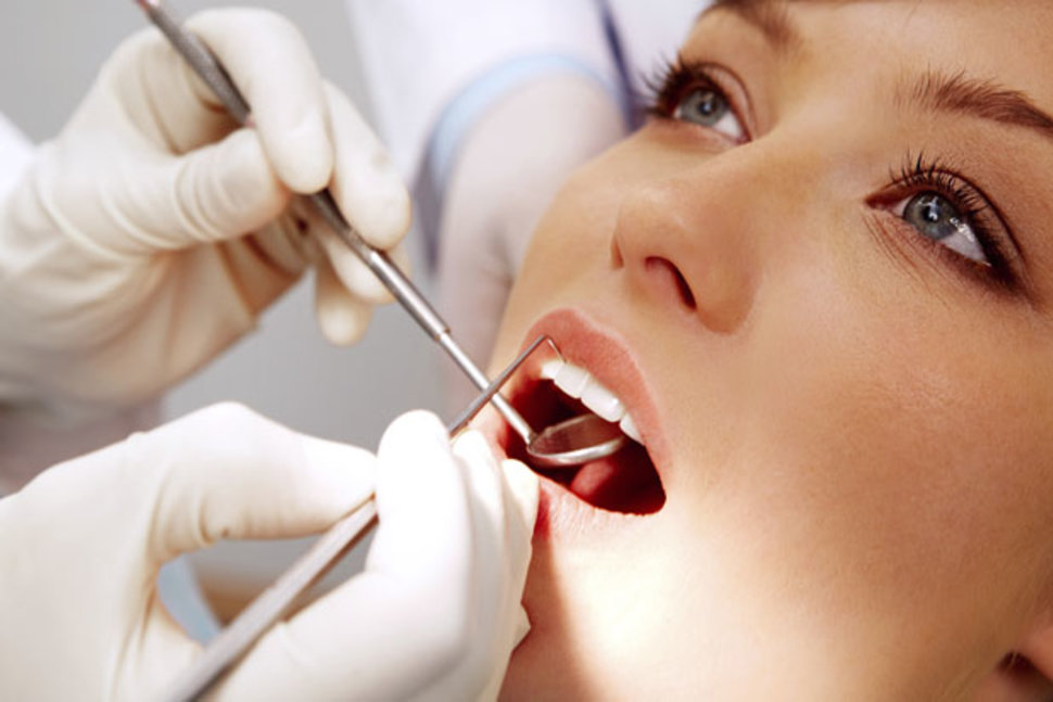
Most patients go to the dentist because of a toothache or any other problem related to their teeth, but they are not the only objects of treatment in dentistry. The fact is that the maxillofacial region is capable of presenting many unpleasant surprises associated with diseases of the neck, mucous and soft tissues of the oral cavity. You may experience an inflammatory process that will be difficult to tie to your teeth, but they may be the likely cause of the disease. Thus, knowing in advance the signs of inflammatory processes, you will be able to respond to the situation in time and not bring the disease to a chronic form by contacting a specialist for treatment.
The reasons
The most likely cause of a jaw abscess is mechanical damage, trauma, or periodontal pockets (gaps between the tooth and gum that can become infected). An abscess can be caused by any infection that has entered the damaged area both from the outside and through the body's bloodstream. If a patient has chronic tonsillitis, streptococci and staphylococci, which constantly multiply in hypertrophied palatine tonsils, can be the cause of inflammation. In this case, the patient is recommended not only to treat the abscess itself and damaged soft tissues of the oral cavity, but also to remove the tonsils if their treatment is not possible. Otherwise, infection may recur repeatedly.
Symptoms and signs
To determine the presence of an inflammatory process, it is enough to know a number of common signs inherent in this disease:
- persistent severe headaches, general malaise, chills;
- in some cases, an increase in body temperature, in particular hyperemia of the inflamed area;
- leukocytosis;
- the presence of fluctuation (accumulation of pus) under the mucosa in the form of a small reddened swelling.
If the above signs are present, the patient is advised to immediately consult a doctor for prompt treatment, otherwise the inflammation may intensify, grow into neighboring areas, develop into more serious diseases or give complications to the respiratory system.
Based on the presence of the upper and lower parts of the jaw in a person, these inflammatory processes can be divided into two types: abscess of the lower jaw (the abscess of the submandibular can also be attributed to the same type, since their sources of origin are the same) and the upper jaw.
Maxillary abscess
The most common source of infection is the upper wisdom teeth. Causes difficulty in opening the mouth and swallowing.
Mandibular abscess
Most often, the infection spreads from the lower large molars (molars and premolars). The patient's complaints are mostly associated with pain when chewing and swallowing.
A submandibular abscess is characterized by visually noticeable and painful swelling in the submandibular triangle, and the shape of the face may be distorted.
Treatment and prevention
Treatment of a jaw abscess consists in opening the abscess and draining the fluid, after which the damaged area is disinfected. In case of high temperature, the patient is prescribed antibiotics, with a general weakening of the immune status - immunomodulatory drugs, recommendations for taking analgesics are also given by the doctor. In rare cases, for better healing of the postoperative incision, physiotherapy procedures, UVI are prescribed.
To prevent inflammation of this kind, it is advisable to visit the dentist every six months, heal periodontal pockets on time, adhere to a sparing diet enriched with vitamins, and also use appropriate therapeutic toothpastes.
Some adherents of alternative medicine believe that the above inflammations of the maxillofacial region can be easily cured without resorting to surgery. Of course, there is a possibility that the abscess will open on its own, however, if it is not cleaned and the remnants of dead particles and pathogenic bacteria are not removed from the wound, there will be a high probability of an acute state becoming chronic or phlegmon, as well as intoxication of the body with decay products remaining in the untreated abscess .
Abscess of the maxillofacial region occurs due to damage or inflammation of the skin of the face, oral mucosa, lips, nose, eyelids. Less commonly, abscesses occur due to the spread of infection from the odontogenic focus. In the absence of treatment of abscesses, purulent decay and purulent fusion of adjacent tissues begin.
Etiology and pathogenesis. An abscess is caused by streptococcal and staphylococcal microflora, the most common cause is dental disease and inflammation in the maxillofacial zone. Furunculosis, tonsillitis, tonsillitis in chronic course are complicated by maxillary abscesses. Damage to the skin and mucous membranes in the mouth area, infection during dental procedures can provoke an abscess of the maxillary zone.
General infectious diseases proceeding according to the type of sepsis, as a result of the spread of microorganisms by blood and lymph, cause multiple abscesses in various organs and tissues, including abscesses of the maxillary zone.
An abscess of the maxillofacial region may occur due to trauma to the face. During military operations and natural disasters, due to the lack of first aid, dislocations and fractures of the jaws are often complicated by abscesses.
Perioapical and pericoronal foci of inflammation and periodontal pockets during exacerbations can provoke a jaw abscess due to bone resorption.
clinical picture. The formation of an abscess is preceded by toothache as in periodontitis. Biting in the affected area increases pain. Further, a dense edema joins with the formation of a painful seal. For an abscess that develops under the mucous membrane, bright hyperemia and protrusion of the affected focus are characteristic. Sometimes facial asymmetry is noted.
In the absence of therapy, the general condition of the patient worsens: the body temperature rises, food is refused. After spontaneous opening of the abscess, the pain subsides, the contours of the face take on normal outlines, and the general state of health stabilizes. But due to favorable conditions for microorganisms in the oral cavity, the process becomes chronic, so its spontaneous opening does not indicate a cure. With short-term weakening of the immune system, perimaxillary abscesses become aggravated. Chronic suppuration from fistulous passages is possible, it is accompanied by bad breath and ingestion of purulent masses. There is a sensitization of the body by decay products, allergic diseases are exacerbated.
Abscesses of the floor of the mouth are characterized by hyperemia in the sublingual zone with the rapid formation of an infiltrate. Conversation and eating become sharply painful, hypersalivation is noted. The mobility of the tongue decreases, it rises slightly upward so as not to come into contact with the emerging abscess. As the swelling increases, the general condition worsens. With spontaneous opening, pus spreads to the peripharyngeal region and neck, which leads to the emergence of secondary purulent foci.
The abscess of the palate often occurs as a complication of periodontitis of the upper second incisor, canine and second premolar. During the formation of an abscess, there is hyperemia and soreness of the hard palate, after bulging, the pain becomes more intense, food intake is difficult. With spontaneous opening, purulent contents spread to the entire area of the hard palate with the development of osteomyelitis of the palatine plate.
If a cheek abscess occurs, then, depending on the location and depth, swelling and redness may be more pronounced on the outside or on the side of the oral mucosa. The soreness of the focus is moderate, with the work of facial muscles, the pain intensifies. The general condition practically does not suffer, but the abscess of the cheek is dangerous by spreading to neighboring parts of the face even before the opening of the abscess.
Abscess of the tongue begins with soreness in the thickness of the tongue, the tongue increases in volume, becomes inactive. Speech, chewing and swallowing of food are sharply difficult and painful. Sometimes a feeling of suffocation can occur with an abscess.
In the focus of inflammation, an infiltrate is formed, in the area of \u200b\u200bwhich the skin or mucous membrane is hyperemic, tense. Fluctuation is determined in the center of the infiltrate. The boundaries of the altered tissues are clearly defined. Often the skin or mucous membrane in the area of the abscess bulges above the surface.
For a correct prognosis and timely subsequent therapy, it is necessary to differentiate an abscess from a furuncle, abscessing lymphadenitis and festering atheroma or congenital cyst.
Treatment. Superficial abscesses on the face in children of older age groups can be opened under local anesthesia. It must be remembered that infiltration of inflamed tissues with an anesthetic causes severe pain. Deep tissue abscesses and abscesses in children of younger age groups should be opened under general anesthesia. It is necessary to carefully evaluate the topography of the abscess in relation to the surrounding tissues, since pronounced reactive edema and an abundance of adipose tissue "mask" the true location of the abscess. For the correct choice of the incision site, this factor must be taken into account. In the presence of an abscess, the depth of the incision should not exceed the thickness of the skin. The subsequent opening of the abscess is achieved by advancing a closed clamp of the "Mosquito" type to the cavity of the abscess. After the appearance of the first portion of pus, the jaws of the clamp are moved apart, and the cavity is emptied. Drainage is introduced into the latter.
The formation of an inflammatory purulent focus in the tissues of the maxillofacial zone of the face. It is manifested by local swelling, redness and fluctuation (swelling) of the skin over the focus of inflammation, facial asymmetry, difficulty and pain in swallowing, and intoxication phenomena. It can develop into diffuse inflammation - phlegmon, with involvement in the process of the peripharyngeal and infraorbital region, neck. Treatment is always surgical - opening and draining the abscess cavity.
General information
- This is a limited focus of purulent inflammation of the tissues of the maxillofacial zone. In the absence of treatment of abscesses, purulent decay and purulent fusion of adjacent tissues begin.
Causes of the maxillary abscess
An abscess is caused by streptococcal and staphylococcal microflora, the most common cause is dental disease and inflammation in the maxillofacial zone. Furunculosis, tonsillitis, tonsillitis in chronic course are complicated by maxillary abscesses. Damage to the skin and mucous membranes in the mouth area, infection during dental procedures can provoke an abscess of the maxillary zone.
General infectious diseases proceeding according to the type of sepsis, as a result of the spread of microorganisms by blood and lymph, cause multiple abscesses in various organs and tissues, including abscesses of the maxillary zone. Abscess of the maxillary zone may occur due to facial trauma. During military operations and natural disasters, due to the lack of first aid, dislocations and fractures of the jaws are often complicated by abscesses. Perioapical and pericoronal foci of inflammation and periodontal pockets during exacerbations can provoke a jaw abscess due to bone resorption.
Symptoms of a maxillary abscess
 The formation of an abscess is preceded by toothache as in periodontitis. Biting in the affected area increases pain. Further, a dense edema joins with the formation of a painful seal. For an abscess that develops under the mucous membrane, bright hyperemia and protrusion of the affected focus are characteristic. Sometimes facial asymmetry is noted.
In the absence of therapy, the general condition of the patient worsens: the body temperature rises, food is refused. After spontaneous opening of the abscess, the pain subsides, the contours of the face take on normal outlines, and the general state of health stabilizes. But due to favorable conditions for microorganisms in the oral cavity, the process becomes chronic, so its spontaneous opening does not indicate a cure. With short-term weakening of the immune system, perimaxillary abscesses become aggravated. Chronic suppuration from fistulous passages is possible, it is accompanied by bad breath and ingestion of purulent masses. There is a sensitization of the body by decay products, allergic diseases are exacerbated.
Abscesses of the floor of the mouth are characterized by hyperemia in the sublingual zone with the rapid formation of an infiltrate. Conversation and eating become sharply painful, hypersalivation is noted. The mobility of the tongue decreases, it rises slightly upward so as not to come into contact with the emerging abscess. As the swelling increases, the general condition worsens. With spontaneous opening, pus spreads to the peripharyngeal region and neck, which leads to the emergence of secondary purulent foci.
The abscess of the palate often occurs as a complication of periodontitis of the upper second incisor, canine and second premolar. During the formation of an abscess, there is hyperemia and soreness of the hard palate, after bulging, the pain becomes more intense, food intake is difficult. With spontaneous opening, purulent contents spread to the entire area of the hard palate with the development of osteomyelitis of the palatine plate.
If a cheek abscess occurs, then, depending on the location and depth, swelling and redness may be more pronounced on the outside or on the side of the oral mucosa. The soreness of the focus is moderate, with the work of facial muscles, the pain intensifies. The general condition practically does not suffer, but the abscess of the cheek is dangerous by spreading to neighboring parts of the face even before the opening of the abscess.
Abscess of the tongue begins with soreness in the thickness of the tongue, the tongue increases in volume, becomes inactive. Speech, chewing and swallowing of food are sharply difficult and painful. Sometimes a feeling of suffocation can occur with an abscess.
Diagnosis and treatment of maxillary abscess
The diagnosis is made on the basis of a visual examination of the dentist and patient complaints. Sometimes during the survey it turns out that there were boils of the facial zone, there are chronic infectious diseases. Before a visit to the doctor, it is recommended to take analgesics, rinse the mouth with antiseptic solutions, self-administration of antibiotics is unacceptable. The ultimate goal of treatment is the complete elimination of the infectious process and the restoration of impaired functions in the shortest possible time.
The treatment regimen depends on the stage of the disease, on the virulence of the microorganism and on the characteristics of the response from the macroorganism. Localization of abscesses of the maxillary zone, the age of the patient and the presence of concomitant diseases significantly affect the principles of treatment. The more complicating factors, the more intensive the therapy should be.
During the period of treatment of abscesses of the maxillary zone, it is recommended to follow a diet with a predominance of pureed soups and mashed potatoes. If there is a persistent refusal of food, they resort to intravenous administration of protein solutions. In the presence of a formed abscess, its opening is shown, followed by drainage of the cavity. In other cases, they resort to antibiotic therapy, and only if it is inappropriate, the question of surgical treatment is raised.
Antibiotics are prescribed in the form of injections or in tablet forms, an additional course of vitamin therapy is carried out. Immunostimulants and detoxification therapy are shown. Rinsing the mouth with warm solutions of furacilin and soda relieves swelling and prevents the spread of infection. In the presence of a pronounced pain syndrome, analgesics are used. With complex therapy started on time, the prognosis is usually favorable, recovery occurs within 6-14 days.
Phlegmon of the submandibular region is an accumulation of purulent substances that affect fatty tissue in the appropriate place. Pathology progresses rather quickly and spreads to bone, muscle tissue, and tendons. The affected areas are hot, reddened, cause pain when pressed.
It differs from the simple submandibular region in the specific blurring of the boundaries and the involvement of adjacent tissues.
Varieties of pathology
Taking into account how and for what reasons the disease developed, it can be primary and secondary. The first variant of the disease is an independent pathology, which appears due to the ingestion of pathogenic microorganisms. Bacteria are activated against the background of a weakened immune system. The secondary form of the disease manifests itself as a result of the spread of pus through the soft tissues of the internal systems due to an abscess, a bursting boil and other accumulations.
In addition, phlegmon of the submandibular region is acute and chronic. The first is characterized by a sharp deterioration in the patient's well-being, an increase in body temperature up to 40 degrees. Chronic pathology has a sluggish character without obvious changes. At the same time, the surface of the inflamed area noticeably turns blue and thickens.
Code of phlegmon of the submandibular region according to ICD-10 (International Classification of Diseases) - K12.2. Pathology is also deep and superficial. The first form is characterized by inflammation of nearby tissues under the layers of the epithelium. With a superficial disease, the soft organs located under the muscles are affected.
By the way, this pathology can develop not only in the jaw area, but also in any other part of the body.
Kinds
Specialists distinguish several types of phlegmon of the submandibular region:
- Serous. This is the initial stage of the disease. Exudate accumulates in injured areas, and adipose tissue is infiltrated. Cellulose in its structure resembles jelly. The boundary between healthy and affected tissues is not clear.
- Purulent. This stage is distinguished by histolysis - the process of tissue melting with the formation of pus in the future. In this case, the exudate becomes whitish, yellow or greenish, cloudy. There are fistulas and sores. In the case of the spread of the inflammatory process, the pathology covers the muscle and bone tissue, which is also subsequently injured.
- Putrefactive. This form is accompanied by the production of foul-smelling gases. The tissues become loose, similar to a dark semi-liquid mass. Putrid phlegmon is necessarily accompanied by severe intoxication.
- Necrotic. With this form, necrotic foci appear, which subsequently melt and are rejected. As a result, instead of them, wounds appear. In the case of a favorable course of pathology, the inflamed area is separated and subjected to an abscess, which is easily opened.
- Anaerobic. It is a serous disease with widespread necrotic lesions. This form is characterized by the formation of gas bubbles. Injured tissues become gray, acquire a terrible smell. When pressing on the injured area, you can hear a crunch that occurs due to gases.
All stages of phlegmon of the submandibular region (ICD-10 code above) have an acute course and often become malignant.
Pathogen
A direct prerequisite for the development of pathology are pathogenic microorganisms. Leaking through the wound, they penetrate the lymph and blood, after which they spread throughout the body. As a rule, Staphylococcus aureus and Streptococcus aureus become the causative agents of phlegmon of the submandibular region. However, the reasons may lie in the vigorous activity of other bacteria:
- Pseudomonas aeruginosa;
- clostridia;
- proteus;
- peptococcus;
- coli;
- pneumococcus;
- paratyphoid or diphtheria bacillus.
Microorganisms usually enter the tissue through open wounds.
In some cases, through biological fluids, bacteria penetrate into fatty tissues from an infectious source already present in the body. A pathogenic focus can occur against the background of tonsillitis, furunculosis, as well as other diseases of the oral, nasal cavity, and larynx.
It is likely that the infection will spread to nearby tissues due to a breakthrough. Sometimes a phlegmon of the submandibular region develops after various chemical compounds, such as kerosene or turpentine, enter the subcutaneous tissue.
Etiology
The progression of the inflammatory phenomenon usually begins with periadenitis or adenitis, much less often as a consequence of the transition of infection from nearby tissues or osteomyelitis of the lower jaw. As a rule, submandibular phlegmon is a consequence of odontogenic infections of the anterior teeth.
In other words, various complications of dental ailments are quite capable of leading to the development of this disease. It is the odontogenic phlegmon of the submandibular region that is considered the most common type of disease with a specific location.
Causes of pathology
Most often, its appearance is associated with difficulty in the eruption of wisdom teeth or is the result of a complicated course of periostitis, lymphadenitis and osteomyelitis. However, the list of causes of phlegmon of the submandibular region is not limited to this. There are a number of additional factors.
The risk of phlegmon is significantly increased if a person suffers from such diseases:
- tuberculosis:
- diabetes;
- immunodeficiency;
- blood problems, such as thrombocytopenia or anemia;
- addiction to alcohol or drugs.
Signs of an inflammatory process
- increase in body temperature up to 39-40 degrees;
- nausea and vomiting;
- noticeable lethargy, decreased performance;
- migraine;
- heart rhythm disturbances;
- strong thirst;
- intoxication;
- decrease in the amount of urine during bladder emptying.

Clinical picture
The submandibular triangle loses its features, painful swelling occurs. The pathological area noticeably reddens and swells. Lymph nodes located near the affected area increase significantly. In the case of unilateral pathology, this usually affects only one side. For example, in a patient with odontogenic phlegmon of the submandibular region on the left, as a rule, the lymph node increases only on this side. The injured area tactilely seems hot, the epithelium on it is shiny. The pain syndrome is shown at the movement.
As the pathological condition progresses, hyperemia occurs, the tension constantly increases, the skin ceases to fold. Palpation becomes more painful each time. There is collateral edema. It is painful for the patient to open his mouth, while the jaws can be reduced in different ways. In some cases, swallowing is also accompanied by pain. A terrible smell comes from the oral cavity, there is excessive production of saliva. The face can be distorted, the tissues become puffy in the neck and chin area. The general well-being of the patient is determined by the virulence of the infection.
Superficial pathology is detected with ease. A qualified specialist can easily recognize this disease during a visual examination. But the deep forms of phlegmon of the submandibular region require additional research:
- radiography;
- puncture of the injured area;
- ultrasound examination;
- magnetic resonance or computed tomography.

Possible complications
Progressing and spreading throughout the body, pathogenic bacteria can cause other diseases:
- faces;
- purulent meningitis;
- thrombophlebitis;
- blood poisoning;
- lymphadenitis.
If the pathological process covers nearby tissues, the lungs and joints are injured, osteomyelitis appears.
The most dangerous consequence of the disease is purulent arteritis. With this disease, the walls of blood vessels are affected, as a result of which extensive bleeding opens.
Treatment of phlegmon of the submandibular region
This disease threatens an infected person with a fatal outcome, so therapy is carried out exclusively in stationary conditions. In the initial stages of the pathology, the patient can do without surgical intervention. A patient with a confirmed diagnosis of "phlegmon of the submandibular region" is prescribed:
- warming manipulations using infrared radiation, compresses and heating pads;
- special compresses with mercury ointment, but this method cannot be used with UHF.
Surgical intervention
If an infiltrate has already formed on the inflamed area, then the operation is necessary, especially at the purulent stage of the pathology. The intervention is performed under general anesthesia. The doctor makes a large incision covering the deep and superficial layers of the epithelium.

After removing the pus, the remaining wound is treated with water and disinfectants. Drainage is made by means of tubes, rubber graduates and semi-tubes. After surgery, patients feel much better.
After the manipulations, a compress with Levomekol ointment and hypertonic solution is applied to the wound.
rehabilitation period
Patients should keep in mind that it is impossible to use dressings with the addition of tetracycline ointment or liniment according to Vishnevsky immediately after the operation. After all, these drugs interfere with the necessary outflow of pus. To speed up the process of rejection of damaged cells, you can use necrolytic drugs such as Terrilitin or Trypsin.
After removing the contents of the inflamed area, therapeutic gauze dressings are applied.
For faster healing, you can take Troxevasin ointment or methyluracil. To improve local immunity, fatty ointments are perfect: streptocid, synthomycin, neomycin. To prevent re-infection, you can use water-based medications: deoxydin ointment or Levosin.
To alleviate the general condition of the patient, other therapeutic procedures can be used:
- To speed up the process of tissue scarring, sea buckthorn or rosehip oil, as well as Troxevasin, are used.
- In the event that the wounds are too deep or do not heal for a long time, dermoplasty is performed.
- In acute phlegmon, the patient must be prescribed antibiotics, the most effective of which in such a situation are "Erythromycin", "Gentamicin", "Cefuroxime". The patient should use these medicines until the inflammation completely disappears.

- At the anaerobic stage, the patient is given
- Calcium chloride solution is used to neutralize toxins and stabilize the acid-base balance of the blood. This substance is also used to tone blood vessels.
- To activate the functioning of the heart muscles, a glucose solution is injected intravenously.
- To maintain immunity, the patient is prescribed vitamin complexes like Alfavit and Vitrum.
Prevention
To prevent the development of such an unpleasant disease as phlegmon of the submandibular region, it is necessary:
- Treat all open wounds with antiseptics.
- In the event of the first signs of pathology, you should immediately contact a dermatologist.
- You need to visit the dentist twice a year.
- Avoid skin contact with household chemicals and penetration of aggressive chemicals into its deep layers.