Meiosis morphology of the main phases of meiosis. Definition and types of meiosis
Meiosis, the most important process of cell division, which occurs on the eve of the formation of germ cells and was discovered at the end of the 19th century, has long been the subject of close attention of a very narrow circle of cytologists. It came to the attention of molecular biologists only in the 1990s. The rapid development of research in this area was facilitated by work on the molecular genetics of model objects, as well as the emergence of new immunocytochemical methods, which gave researchers a convenient way to study proteins involved in meiosis.
In all eukaryotes, during meiosis, a submicroscopic structure is formed, called synaptonemal complex(from Greek synaptos - connected, peta - thread). The study of the molecular organization of this complex and its role in meiosis showed that it is needed for the recombination of chromosomes and the reduction of their number. This will be discussed in this article.
But first, let us recall the basic information about meiosis, which consists of two divisions: meiosis I and meiosis II. As a result of reduction division (meiosis I), the number of chromosomes in daughter cells is halved compared to the set of chromosomes in the parent cell. This is because the amount of DNA in chromosomes doubles only once before meiosis I (Fig. 1). A twofold reduction in the number of chromosomes during the formation of germ cells makes it possible to restore the initial (diploid) number of chromosomes during fertilization and maintain its constancy. This requires a strict separation of pairs of homologous chromosomes between germ cells. With errors, aneuploidy occurs - a lack or excess of chromosomes, and this imbalance leads to the death of the embryo or severe developmental anomalies (in humans, to the so-called chromosomal diseases).
Structure and function of the synaptonemal complex
The synaptonemal complex consists of two protein axes of homologous chromosomes connected by a protein zipper (Fig. 2). Zipper prongs are rod-shaped dimers of parallel-laid and identically oriented protein molecules with a long α-helix in the middle of the molecule. Yeast S. cerevisiae - it is the Zip1 protein, in mammals and humans it is SCP1 (SYCP1). These proteins are attached with their C-termini to the chromosome axes (lateral elements of the complex), while their N-termini are directed towards each other, inside the central space (Fig. 3). At the N-terminus of the molecules there are charged "spurs" - alternating peaks in the densities of positive and negative charges of amino acids (Fig. 4), the complementary interaction of which provides a strong electrostatic connection of the teeth.
The so-called central space of the complex (the gap between the protein axes, filled with the teeth of the "fastener", about 100 nm wide), as well as the entire complex (its cross section is about 150-200 nm) are not visible in a conventional light microscope, since the entire complex is masked by chromatin. For the first time, the synaptonemal complex was seen on ultrathin (0.8 µm thick) sections of the testes of crayfish and mice using a transmission electron microscope. It was discovered in 1956 independently by two American researchers - M. Moses and D.V. Fossett.
Now, when studying the complex, the so-called microspreading method is used. Testis cells (or plant anthers) after hypotonic shock are placed on a plastic substrate deposited on a glass slide. The content of the burst cell is fixed with a weak solution of formaldehyde and contrasted with salts of heavy metals (best of all - AgNO 3). The glass is examined in a phase-contrast microscope and, by indirect signs, cells are selected that should contain the complex. A circle of film with the desired cell is picked up on a metal mesh and placed in an electron microscope (Fig. 5). If necessary, before contrasting cells are treated with antibodies to proteins of interest to the researcher. These antibodies are labeled with calibrated colloidal gold beads, which are clearly visible under an electron microscope.
During prophase I of meiosis, the synaptonemal complex retains parallel homologous chromosomes almost until they are built on the equator of the cell (metaphase I). Chromosomes are connected using the synaptonemal complex for some time (from 2 hours in yeast to 2-3 days in humans), during which homologous DNA regions are exchanged between homologous chromosomes - crossing over. In crossing over, which occurs with a frequency of at least one event (more often two, less often three or four) per pair of homologous chromosomes, dozens of meiosis-specific enzyme proteins participate.
The molecular mechanism of crossing over and its genetic implications are two big topics that are beyond the scope of this story. We are interested in this process because, as a result of it, homologous chromosomes are firmly bound by crossed DNA molecules (chiasmata) and the need for pairwise retention of the chromosome with the help of the synaptonemal complex disappears (after crossing over, the complex disappears). Homologous chromosomes connected by chiasmata line up at the equator of the cell division spindle and diverge with the help of the cell division spindle threads into different cells. After meiosis is completed, the number of chromosomes in daughter cells is halved.
So, only on the eve of meiosis I, the structure of chromosomes changes radically. A very specific intranuclear and interchromosomal structure - the synaptonemal complex - occurs once in the life cycle of an organism for a short time for pairing of homologous chromosomes and crossing over, and then is dismantled. These and many other events during meiosis at the molecular and subcellular (ultrastructural) levels are provided by the work of numerous proteins that perform structural, catalytic, and kinetic (motor) functions.
Proteins of the synaptonemal complex
Back in the distant 1970s, we obtained indirect evidence that the synaptonemal complex is formed by self-assembly of its elements, which can occur even in the absence of chromosomes. The experiment was set by nature itself, and we managed to observe it. It turned out that in the porcine roundworm, in the cytoplasm of cells preparing for meiosis I, packets or "stacks" of absolutely correctly stacked morphological elements of the synaptonemal complex appear (although there are no chromosomes in the cytoplasm: they are in the nucleus). Since there is no synaptonemal complex in the cell nuclei at the stage of preparation of cells for meiosis, it has been suggested that the control of the sequence of meiotic events in this primitive organism is imperfect. An excess of newly synthesized proteins in the cytoplasm leads to their polymerization and the appearance of a structure that does not differ from the synaptonemal complex. This hypothesis was confirmed only in 2005 thanks to the work of an international group of researchers working in Germany and Sweden. They showed that if the gene encoding the mammalian zipper prong protein (SCP1) is introduced into somatic cells growing on an artificial nutrient medium and activated, a powerful network of SCP1 proteins “zipped” between itself in the same way as in the central space of the complex. The formation of a layer of continuous protein "zippers" in cell culture means that the ability of the proteins of the complex to self-assemble, which we predicted, has been proven.
In 1989 and in 2001 Our laboratory staff O. L. Kolomiets and Yu. S. Fedotova investigated the natural “dismantling” of synaptonemal complexes at the final stages of their existence. This multi-step process has been best observed in pollen mother cells in rye anthers, where there is partial synchrony of meiosis. It turned out that the lateral elements of the complex are dismantled by gradual “unwinding” of the protein supercoil, which has three levels of packaging (Fig. 6).
The basis of the extended lateral elements is a complex of four cohesin proteins (from the English. cohesion- grip). On the eve of meiosis, a specific Rec8 cohesin protein appears in the chromosomes, which replaces the somatic cohesin Rad21. Then three other cohesin proteins, which are also present in somatic cells, join it, but instead of the somatic cohesin SMC1, the meiosis-specific protein SMC1b appears (its N-terminus differs by 50% from the N-terminus of the somatic SMC1 protein). This cohesin complex sits within the chromosome between two sister chromatids, holding them together. Meiosis-specific proteins bind to the cohesin complex, which become major proteins of chromosome axes and turn them (these axes) into lateral elements of the synaptonemal complex. In mammals, the major proteins of the synaptonemal complex are SCP2 and SCP3, in yeast, the Hop1 and Red1 proteins, and the meiosis-specific protein, Rec8.
The evolutionary paradox of proteins
In mammals and yeast, the proteins of the synaptonemal complex have different amino acid sequences, but their secondary and tertiary structures are the same. Thus, the zipper protein SCP1 in mammals and the non-homologous protein Zip1 in yeast are built according to a single plan. They consist of three amino acid domains: the central one is an α-helix capable of forming a second-order helix (supercoiling), and two terminal domains are globules. The major proteins SCP2 and SCP3, which have no homology with the Hop1 and Red1 proteins of yeast and, apparently, with the still insufficiently studied proteins of the complex in plants, also build morphologically and functionally identical structures of the synaptonemal complex. This means that the primary structure (sequence of amino acids) of these proteins is an evolutionarily neutral trait.
So, non-homologous proteins in evolutionarily distant organisms build the synaptonemal complex according to a single plan. Explaining this phenomenon, I will use the analogy with the construction of houses from different materials, but according to a single plan. It is important that such houses have walls, ceilings, a roof, and that building materials meet the conditions of strength. Similarly, the formation of the synaptonemal complex requires lateral elements ("walls"), transverse filaments ("zipper teeth") - "overlays" and a central space (room for the "kitchen"). “Kitchen robots” should fit there - complexes of recombination enzymes assembled into so-called “recombination knots”.
The width of the central space of the synaptonemal complex in yeast, maize, and humans is approximately 100 nm. This is due to the length of single-stranded DNA regions coated with the Rad51 recombination protein. This protein belongs to a group of enzymes (similar to the bacterial recombination protein RecA) that have retained homology since the advent of DNA recombination (about 3.5 billion years ago). The inevitability of the homology of recombination proteins in distant organisms is determined by their function: they interact with the double helix of DNA (the same in bacteria and mammals), dividing it into single strands, cover them with a protein sheath, transfer one strand to the homologous chromosome and restore the double helix there again. Naturally, most of the enzymes involved in these processes retain their homology for more than 3 billion years. In contrast, the synaptonemal complexes that appeared in eukaryotes after the onset of meiosis (about 850 million years ago) are built from non-homologous proteins ... but the scheme of their domain structure is the same. Where did this scheme come from?
A clue is the mentioned Rec8 protein, which begins the formation of chromosome axes in the meiotic cycle and which is present in all studied organisms. It can be assumed that the building material for the axes of meiotic chromosomes and the lateral elements of the synaptonemal complex can be any intermediate proteins that are able to form a fibrous structure (SCP2, Hop1, etc.), interact with the Rec8 cohesin and "settle" on it, like concrete on a metal fittings.
In recent years, experiencing difficulties in conducting experimental work due to insufficient funding, we began to actively use bioinformatics methods. We were interested in the zipper protein in Drosophila. Considering the similarity of the secondary and tertiary structures of yeast Zip1 proteins and human SCP1, we hypothesized that the Drosophila zipper protein has the same structure. We started our work in 2001, when the Drosophila genome had already been sequenced and it became known that it contained approximately 13,000 potential genes. How to find the gene for the protein we are looking for?
Among the 125 meiosis genes known by that time in Drosophila, we foresaw only one candidate for this role. The fact is that the mutation of the gene c(3)G deprived the chromosomes of the ability to connect in pairs with the help of a "zipper" and enter into recombination. We hypothesized that the mutants have a defective protein that forms submicroscopic teeth of the “fastener”. The secondary structure and conformation of the desired protein should be similar to the Zip1 and SCP1 proteins.
Knowing that the gene c(3)G is located on chromosome 3 in Drosophila, we searched the database of this region (comprising 700 kb) for an open reading frame that could encode a similar protein. We understood that in the absence of homology in the primary structure of the desired protein and yeast, their size, organization (of three domains) and the ability of the central domain to form an α-helix of a certain length (about 40 nm) should be similar. This was evidenced by the similarity of the electron microscopic picture of the synaptonemal complex in meiosis in yeast and Drosophila.
Open reading frames were scanned for nearly 80 genes in the search area. Using computer programs that allow predicting the secondary structure of a virtual protein, its physicochemical properties and the distribution of electrostatic charges in molecules, T. M. Grishaeva found such a reading frame at the border of the gene localization zone c(3)G.(This was not very accurately predicted by Japanese geneticists on a microscopic map of chromosomes.) It turned out to be a gene CG1J604 according to the genomic map of the Celera company.
We concluded that this virtual gene must be a long-known gene c(3)G and encode a protein similar to the yeast Zip1 protein. In response to our message, we received an email from S. Hawley from the USA. He experimentally proved that c(3)G encodes a protein that forms a "zipper" between chromosomes during meiosis in Drosophila. The results of our work coincided, but the experimental work of Hawley's group took about seven years, and our computer work by three people took only about three months. The articles went out of print at the same time. In 2003 we published the method of our computer searches and provided examples of similar virtual proteins in other organisms. This work is now readily cited by foreign colleagues, and our method works successfully in their hands in combination with experimental verification. So, in 2005, a group of English biologists discovered the gene and protein of the zipper teeth in the plant Arabidopsis thaliana .
In conclusion, I will give an example of another finding in the field of molecular biology of meiosis, but we must start with mitosis. In order for the chromatids to separate in the anaphase of mitosis, it is necessary to destroy the cohesin that “glues” them together. Hydrolysis of cohesins during mitosis is a genetically programmed event. But in the metaphase of meiosis I, when the homologous chromosomes are lined up at the equator of the cell and the protein spindle is ready to pull them to the poles, the hydrolysis of cohesins is impossible. That is why both chromatids of each chromosome, glued together in the region of the kinetic center of chromosomes (kinetochore), go to the same pole (see Fig. 1). In the late 1990s, Japanese researchers studying meiosis in yeast found that in the kinetochore region, cohesins are protected by a protein they called shugoshin (the root of this term is taken from the samurai lexicon and means protection). Very quickly, the world community of meiosis researchers came to the conclusion that Drosophila, corn, and other objects have similar shugoshin proteins. At the same time, genes that "prohibit" the separation of chromatids in meiosis I in Drosophila were known 10 years before, but their protein product was not deciphered. And in 2005, a group of American researchers from the University of California at Berkeley, including our compatriot and my longtime colleague in the study of meiosis I. N. Golubovskaya, reported that during metaphase I of meiosis in the chromosomes of corn, the ZmSGO1 shugoshin is located on both sides of kinetochores, and it appears in this region only if there is already a Rec8 cohesin, which it protects from hydrolysis (but only in meiosis I). These results were obtained using fluorescent antibodies to proteins and a confocal microscope. It remains to add that Japanese researchers immediately reported that shugoshin protected Rec8 from hydrolysis if shugoshin was dephosphorylated. Phosphorylation and dephosphorylation, as well as acetylation and deacetylation, are important modifications that change the properties of protein molecules.
Applied aspect
Everything told is a beautiful fundamental science, but can this knowledge be used for practical purposes? Can. Back in the mid-1980s, British researchers and our laboratory proved using various experimental models that, using microspreads of synaptonemal complexes, it is possible to detect twice as many chromosomal rearrangements (deletions, translocations, inversions) as compared with the traditional method of analyzing chromosomes at the metaphase stage. (Fig. 7). The fact is that the synaptonemal complex is the skeletal structure of meiotic chromosomes in prophase. At this time, the chromosomes are about 10 times longer, which significantly increases the resolution of the analysis. However, it is practically impossible to study prophase chromosomes entangled into a coil, and the rigid skeletal structures of the synaptonemal complex are not afraid of spreading, and, in addition, an electron microscope is able to distinguish miniaberrations that are inaccessible to a light microscope.
We asked ourselves the question: is it possible to establish the cause of sterility in the offspring of irradiated mice by studying not the chromosomes, but the synaptonemal complex? It turned out that in sterile mice that inherited chromosomal translocations from their parents, these rearrangements are detected using the complex in 100% of the cells under study, and with conventional methods of "metaphase" analysis - only in 50% of the cells. A group of Spanish researchers examined more than 1 thousand men suffering from infertility. In a third of them, the cause of infertility could not be previously determined, and the study of the synaptonemal complex from the cells of the testes of these patients allowed half of them to make a diagnosis: the cause of infertility is the absence of the synaptonemal complex, which is why spermatocytes (sperm progenitor cells) do not develop, i.e. they do not develop. e. there was an "arrest" of the process of meiosis and all spermatogenesis. Similar results were obtained by OL Kolomiets together with doctors from Kharkov. The study of the synaptonemal complex in combination with other methods of analysis increases the percentage of detection of the causes of infertility in the examined male patients from 17 to 30%. Some English clinics already in the 90s of the XX century. used these methods extensively. Such diagnostics, of course, requires high theoretical and practical skills of physicians and the use of electron microscopes. Russian laboratories have not yet reached such a level, with the exception of the Institute of General Genetics. N. I. Vavilov Institute of Cytology and Genetics of the Siberian Branch of the Russian Academy of Sciences (Novosibirsk).
One might think that intensive studies of the mechanisms of meiosis will inevitably lead to the application of the acquired knowledge in those areas of biology and medicine that are associated with the fertility of living organisms, including humans. However, the law of applying scientific achievements in practice is unchanged: it is useless to “implement” something by force. Practitioners themselves must follow the achievements of science and use them. This is the approach used by leading pharmaceutical and biotech firms.
From the discovery of meiosis (1885) to the discovery of the synaptonemal complex (1956), approximately 70 years passed, and from 1956 until the discovery of the proteins of the synaptonemal complex (1986), another 30 years. Over the next 20 years, we learned the structure of these proteins, their coding genes, the interaction proteins in the construction and operation of synaptonemal complexes, in particular, their interaction with protein-enzymes of DNA recombination, etc., i.e. more than in the previous 30-year period of descriptive cytological studies. It is possible that it will take no more than two decades to decipher the main molecular mechanisms of meiosis. The history of science, as well as of the whole civilization, is characterized by the "compression of time", the increasing compaction of events and discoveries.
Literature:
- Page S.L., Hawley R.S.// Anna. Rev. Cell Develop. Biol. 2004. V. 20. P. 525-558.
- Moses M.J.//Chromosoma. 2006. V. 115. P. 152-154.
- Bogdanov Yu.F.// Chromosoma. 1977. V. 61. P. 1-21.
- OllingerR. et al.//Moll. Biol. cell. 2005. V. 16. P. 212-217.
- Fedotova Y.S. et al. // Genome. 1989. V. 32. P. 816-823; Kolomiets O.L. and etc.// Biological membranes. 2001. T. 18. S. 230-239.
- Bogdanov Yu.F. et al. // Int. review. Cytol. 2007. V. 257. P. 83-142.
- Bogdanov Yu.F.// Ontogeny. 2004. T. 35. No. 6. C. 415-423.
- Grishaeva T.M. et al.// Drosophila Inform. Serv. 2001. V. 84. P. 84-89.
- Page S.L., Hawley R.S.// Genes Develop. 2001. V. 15. P. 3130-3143.
- Bogdanov Yu.F. et al. // In Silico Biol. 2003. V. 3. P. 173-185.
- Osman K. et al. // Chromosoma. 2006. V. 115. P. 212-219.
- Hamant O., Golubovskaya I. et al.// Curr. Biol. 2005. V. 15. P. 948-954.
- Kalikinskaya E.I. et al. // Mut. Res. 1986. V. 174. P. 59-65.
- Egozcue J. et al.// Hum. Genet. 1983. V. 65. P. 185-188; Carrara R. et al.// Genet. Mol. Biol. 2004. V. 27. P. 477-482.
- Bogdanov Yu.F., Kolomiets O.L. synaptonemal complex. Indicator of meiosis dynamics and chromosome variability. M., 2007.


Meiosis is a special way of dividing eukaryotic cells, in which the initial number of chromosomes is reduced by 2 times (from the ancient Greek "meion" - less - and from "meiosis" - reduction).
Separate phases of meiosis in animals were described by W. Flemming (1882), and in plants by E. Strasburger (1888), and then by the Russian scientist V.I. Belyaev. At the same time (1887) A. Weissman theoretically substantiated the need for meiosis as a mechanism for maintaining a constant number of chromosomes. The first detailed description of meiosis in rabbit oocytes was given by Winiworth (1900).
Although meiosis was discovered more than 100 years ago, the study of meiosis continues to this day. Interest in meiosis increased dramatically in the late 1960s, when it became clear that the same gene-controlled enzymes could be involved in many DNA-related processes. Recently, a number of biologists have been developing an original idea: meiosis in higher organisms serves as a guarantor of the stability of the genetic material, because during meiosis, when pairs of homologous chromosomes are in close contact, DNA strands are checked for accuracy and damage is repaired that affects both strands at once. The study of meiosis linked the methods and interests of two sciences: cytology and genetics. This led to the birth of a new branch of knowledge - cytogenetics, which is now in close contact with molecular biology and genetic engineering.
The biological significance of meiosis lies in the following processes:
1. Due to the reduction in the number of chromosomes as a result of meiosis in a series of generations during sexual reproduction, the constancy of the number of chromosomes is ensured.
2. Independent distribution of chromosomes in the anaphase of the first division ensures the recombination of genes belonging to different linkage groups (located on different chromosomes). The meiotic distribution of chromosomes among daughter cells is called chromosome segregation.
3. Crossing over in prophase I of meiosis ensures the recombination of genes belonging to the same linkage group (located on the same chromosome).
4. The random combination of gametes during fertilization, together with the above processes, contributes to genetic variability.
5. In the process of meiosis, another significant phenomenon occurs. This is the process of activation of RNA synthesis (or transcriptional activity of chromosomes) during prophase (diplotenes), associated with the formation of lampbrush chromosomes (found in animals and some plants).
This reversion of prophase to the interphase state (during mitosis, mRNA synthesis occurs only in interphase) is a specific characteristic of meiosis as a special type of cell division.
It should be noted that in protozoa, a significant variety of meiotic processes is observed.
In accordance with the position in the life cycle, three types of meiosis are distinguished:
Zygote th (initial) meiosis occurs in the zygote, i.e. immediately after fertilization. It is characteristic of organisms whose life cycle is dominated by the haploid phase (ascomycetes, bisidiomycetes, some algae, sporozoans, etc.).
Gametic(terminal) meiosis occurs during the formation of gametes. It is observed in multicellular animals (including humans), as well as among protozoa and some lower plants, in the life cycle of which the diploid phase predominates.
Intermediate(spore) meiosis occurs during spore formation in higher plants, including between the stages of sporophyte (plant) and gametophyte (pollen, embryo sac).
Thus, meiosis is a form of nuclear division, accompanied by a decrease in the number of chromosomes from diploid to haploid and a change in the genetic material. The result of meiosis is the formation of cells with a haploid set of chromosomes (sex cells).
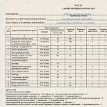
The duration of meiosis may differ depending on the type of plants and animals (Table 1).
Table 1. Duration of meiosis in various plant species
A typical meiosis consists of two consecutive cell divisions, respectively called meiosis I and meiosis II. In the first division, the number of chromosomes is halved, so the first meiotic division is called reduction, less often heterotypic. In the second division, the number of chromosomes does not change; this division is called equational(equalizing), less often - homeotypic. The expressions "meiosis" and "reduction division" are often used interchangeably.
The initial number of chromosomes in meiocytes (cells entering meiosis) is called the diploid chromosome number (2n). The number of chromosomes in cells formed as a result of meiosis is called the haploid chromosome number (n). The minimum number of chromosomes in a cell is called the base number (x). The basic number of chromosomes in a cell corresponds to the minimum amount of genetic information (the minimum amount of DNA), which is called the gene.
The number of genomes in a cell is called the genomic number (n). In most multicellular animals, in all gymnosperms and in many angiosperms, the concept of haploidy-diploidy and the concept of genomic number coincide. For example, in humans n=x=23 and 2n=2x=46.
Morphology of meiosis - characteristics of phases
Interphase
The premeiotic interphase differs from the usual interphase in that the process of DNA replication does not reach the end: approximately 0.2 ... 0.4% of the DNA remains undoubled. Thus, cell division begins at the synthetic stage of the cell cycle. Therefore, meiosis is figuratively called premature mitosis. However, in general, it can be considered that in a diploid cell (2n) the DNA content is 4c.
In the presence of centrioles, they are doubled in such a way that there are two diplosomes in the cell, each of which contains a pair of centrioles.
first division of meiosis
The DNA has been replicated. Prophase I is the longest stage of meiosis.
The prophase I stage is subdivided into the following stages:
leptotena - the stage of thin threads;
zygotene - stage of double threads;
pachytene - the stage of thick threads;
diplotena - crossing over;
diakinesis - the disappearance of the nuclear membrane and nucleolus.
In early prophase (leptoten), preparation for conjugation of chromosomes takes place. The chromosomes are already doubled, but the sister chromatids in them are still indistinguishable. Chromosomes begin to pack (spiralize).
In contrast to the prophase of mitosis, where the chromosomes are located along the nuclear membrane end to end and, being packed, are attracted to the membrane, the leptotene chromosomes with their telomeric regions (ends) are located in one of the poles of the nucleus, forming a “bouquet” figure in animals and squeezing into a ball. synesis" - in plants. Such an arrangement or orientation in the nucleus allows chromosomes to quickly and easily conjugate homologous chromosome loci (Fig. 1).
The central event is the mysterious process of recognition of homologous chromosomes and their pairwise approach to each other occurs in the prophase I zygotene. When conjugation (approach) of homologous chromosomes, pairs are formed - bivalents and the chromosomes are noticeably shortened. From this moment, the formation of the synaptonemal complex (SC) begins. The formation of the synaptonemal complex and the synopsis of chromosomes are synonyms.
Rice. 1. Prophase stage
During the next stage of prophase I - pachytene between homologous chromosomes, close contact is strengthened, which is called synapsis (from the Greek synopsis - connection, connection). Chromosomes at this stage are highly spiralized, which makes it possible to observe them under a microscope.
During synapsis, homologues intertwine, i.e. conjugate. The conjugating bivalents are linked by chiasmata. Each bivalent consists of two chromosomes and four chromatids, where each chromosome comes from its parent. During the formation of synapsis (SC), there is an exchange of sites between homologous chromatids. This process, called crossing over, causes the chromatids to now have a different gene composition.
The synaptonemal complex (SC) in pachytene reaches its maximum development and during this period is a ribbon-like structure located in the space between parallel homologous chromosomes. The SC consists of two parallel lateral elements formed by densely packed proteins and a less dense central element extending between them (Fig. 2).

Rice. 2. Scheme of the synaptonemal complex
Each lateral element is formed by a pair of sister chromatids in the form of a longitudinal axis of the leptoten chromosome and, before becoming part of the SC, is called the axial element. Lateral loops of chromatin lie outside the SC, surrounding it from all sides.
SC development during meiosis:
the leptotene structure of the chromosomes that have entered the leptothene immediately turns out to be unusual: in each homologue, a longitudinal strand is observed along the axis of the chromosomes along its entire length;
zygotene - at this stage, the axial strands of the homologues approach each other, while the ends of the axial strands attached to the nuclear membrane seem to slide along its inner surface towards each other;
pachytene. The SC reaches its greatest development in pachytene, when all its elements acquire maximum density, and chromatin looks like a dense continuous “fur coat” around it.
SC functions:
1. A fully developed synaptonemal complex is necessary for the normal retention of homologues in the bivalent for as long as it is necessary for crossing over and chiasm formation. Chromosomes are connected using the synaptonemal complex for some time (from 2 hours in yeast to 2–3 days in humans), during which homologous DNA regions are exchanged between homologous chromosomes - crossing over (from English, crossing over - cross formation).
2. Prevention of too strong connection of homologues and keeping them at a certain distance, preserving their individuality, creating an opportunity to push off in diplotene and disperse in anaphase.
The process of crossing over is associated with the work of certain enzymes, which, when chiasmata are formed between sister chromatids, “cut” them at the point of intersection, followed by the reunification of the formed fragments. In most cases, these processes do not lead to any disturbances in the genetic structure of homologous chromosomes; there is a correct connection of fragments of chromatids and the restoration of their original structure.
However, another (more rare) variant of events is also possible, which is associated with an erroneous reunion of fragments of cut structures. In this case, there is a mutual exchange of sections of genetic material between conjugating chromatids (genetic recombination).
On fig. Figure 3 shows a simplified diagram of some possible variants of a single or double crossing over involving two chromatids from a pair of homologous chromosomes. It should be emphasized that crossing over is a random event that, with one or another probability, can occur in any region (or in two or more regions) of homologous chromosomes. Consequently, at the stage of maturation of the gametes of a eukaryotic organism in the prophase of the first division of meiosis, the universal principle of random (free) combination (recombination) of the genetic material of homologous chromosomes operates.
In cytological studies of synapsis over the past two decades, an important role has been played by the method of spreading prophase meiotic cells of animals and plants under the action of a hypotonic solution. The method entered cytogenetics after the work of Moses and played the same role that the method of preparing "squashed" preparations for the study of metaphase chromosomes played in its time, saving cytogeneticists from microtome sections.
The Moses method and its modifications have become more convenient than the analysis of SC on ultrathin sections. This method became the basis of meiosis research and gradually covered the issues of gene control of meiosis in animals and plants.

Rice. 3. Separate variants of single and double crossing over involving two chromatids: 1 initial chromatids and a variant without crossing over; 2 single crossing-over in the region A B and crossover chromatids; 3 single crossing over in the B-C region and crossover chromatids; 4 double crossing over and crossover chromatids of several different sites based on the homology of the genetic material of these sites. It is believed that either one of the two sister chromatids of the corresponding chromosome or both chromatids can participate in the conjugation process on each side.
In a dippoten, homologous chromosomes begin to repel each other after mating and crossing over. The process of repulsion begins at the centromere. The divergence of homologues is prevented by chiasma - the junction of non-sister chromatids resulting from the crossing. As the chromatids separate, some of the chiasmata move towards the end of the chromosome arm. Usually there are several crossovers, and the longer the chromosomes, the more there are, therefore, in a diplotene, as a rule, there are several chiasmata in one bivalent.
In the stage of diakinesis, the number of chiasmata decreases. Bivalents are located on the periphery of the nucleus. The nucleolus dissolves, the membrane collapses, and the transition to metaphase I begins. The nucleolus and nuclear membrane are preserved throughout the entire prophase. Before prophase, during the synthetic period of interphase, DNA replication and chromosome reproduction occur. However, this synthesis does not end completely: DNA is synthesized by 99.8%, and proteins - by 75%. DNA synthesis ends in pachytene, proteins - in diplotene.
In metaphase I, the spindle-shaped structure formed by microtubules becomes noticeable. During meiosis, individual microtubules are attached to the centromeres of the chromosomes of each bivalent. Then pairs of chromosomes move to the equatorial plane of the cell, where they line up in a random order. The centromeres of homologous chromosomes are located on opposite sides of the equatorial plane; in the metaphase of mitosis, on the contrary, the centromeres of individual chromosomes are located in the equatorial plane.
In metaphase I, bivalents are located in the center of the cell, in the zone of the equatorial plate (Fig. 4).

Rice. 4. Stages of meiosis: prophase I - metaphase I
Anaphase begins with the separation of homologous chromosomes and their movement towards the poles. In chromosomes without a centromere, attachment cannot exist. In anaphase of mitosis, centromeres divide and identical chromatids separate. In anaphase I of meiosis, the centromeres do not divide, the chromatids remain together, but the homologous chromosomes separate. However, due to the exchange of fragments as a result of crossing over, the chromatids are not identical, as at the beginning of meiosis. In anaphase I, the conjugating homologues diverge towards the poles.
In daughter cells, the number of chromosomes is half as much (haploid set), while the DNA mass is also halved and the chromosomes remain dichromatid. The exact divergence of homologous pairs to opposite poles underlies the reduction of their number.
In telophase I, chromosomes are concentrated at the poles, some of them decondense, due to which the spiralization of chromosomes weakens, they lengthen and again become indistinguishable (Fig. 5). As the telophase gradually passes into interphase, the nuclear envelope (including fragments of the parent cell nucleus envelope) and the cell septum arise from the endoplasmic reticulum. Finally, the nucleolus re-forms and protein synthesis resumes.

Rice. 5. Stages of meiosis: anaphase I - telophase I
In interkinesis, nuclei are formed, each of which contains n dichromatid chromosomes.
The peculiarity of the second division of meiosis is, first of all, that chromatin doubling does not occur in interphase II, therefore, each cell entering prophase II retains the same n2c ratio.
Second division of meiosis
During the second division of meiosis, the sister chromatids of each chromosome diverge towards the poles. Since crossing over could occur in prophase I and sister chromatids could become non-identical, it is customary to say that the second division proceeds according to the type of mitosis, but this is not true mitosis, in which daughter cells normally contain chromosomes identical in shape and set of genes.
At the beginning of the second meiotic division, the chromatids are still connected by centromeres. This division is similar to mitosis: if the nuclear membrane formed in telophase I, now it is destroyed, and by the end of the short prophase II, the nucleolus disappears.

Rice. 6. Stages of meiosis: prophase II-metaphase II
In metaphase II, the spindle and chromosomes, consisting of two chromatids, can again be seen. Chromosomes are attached by centromeres to spindle threads and line up in the equatorial plane (Fig. 6). In anaphase II, the centromeres divide and separate, and sister chromatids, now chromosomes, move toward opposite poles. In telophase II, new nuclear membranes and nucleoli are formed, the contraction of chromosomes weakens, and they become invisible in the interphase nucleus (Fig. 7).

Rice. 7. Stages of meiosis: anaphase II - telophase II
Meiosis ends with the formation of haploid cells - gametes, tetrads of spores - descendants of the original cell with a doubled (haploid) set of chromosomes and haploid DNA mass (original cell 2n, 4c, - spores, gametes - n, c).
The general scheme for the distribution of chromosomes of a homologous pair and the two pairs of differing allelic genes contained in them during two divisions of meiosis is shown in Fig. 8. As can be seen from this scheme, two fundamentally different variants of such a distribution are possible. The first (more probable) variant is associated with the formation of two types of genetically different gametes with chromosomes that have not undergone crossing overs in the regions where the genes under consideration are localized. Such gametes are called non-crossover. In the second (less probable) variant, along with non-crossover gametes, crossover gametes also arise as a result of genetic exchange (genetic recombination) in regions of homologous chromosomes located between the loci of two non-allelic genes.

Rice. 8. Two variants of the distribution of chromosomes of a homologous pair and the non-allelic genes contained in them as a result of two divisions of meiosis
Nikolai Mushkambarov, Dr. biol. Sciences
Humanity is aging, and yet everyone wants to live not just long, but also without those diseases that come with age. Over the past half century, there have been many "revolutionary" theories of aging, almost every one of which offers a sure and reliable way to slow down or even stop time. Every year - new sensations, new discoveries and new statements, encouraging and promising. Peptide bioregulators, longevity elixir, life-giving ions, or SkQ antioxidant. Run to the pharmacy, pay and live, according to the enclosed instructions, up to 100-120 years! To what extent can sensational discoveries be trusted and what is the “truth about aging”?
Professor N. N. Mushkambarov. Photo by Andrey Afanasiev.
August Weismann (1834-1914) German zoologist and evolutionist. He created a theory according to which hereditary traits are preserved and transmitted through the ageless germ plasm.
Leonard Hayflick is an American microbiologist. In the 1960s, he discovered that under laboratory conditions, human and animal cells can divide only a limited number of times.
Alexey Matveyevich Olovnikov is a Russian biochemist. To explain Hayflick's experiments in 1971, he put forward a hypothesis about the shortening of the terminal sections of chromosomes (telomeres) with each cell division.
Science and life // Illustrations
Elizabeth Blackburn and Carol Greider are American biologists. In 1985, the enzyme telomerase was discovered. The mechanism of action of telomerase is the repeated coding of new nucleotide sequences at the terminal sections of telomeres and the restoration of their original length.
Benjamin Gompertz (1779-1865), British mathematician. Proposed a function that describes the statistics of human mortality depending on age. This feature has been used to evaluate risks in life insurance.
The book by M. M. Vilenchik “The Biological Foundations of Aging and Longevity”, published in 1976, was one of the first popular science books on the topic of aging and was a huge success.
Scheme of meiosis (on the example of a pair of homologous chromosomes). In the prophase of the first division of meiosis, the chromosomes double; then homologous chromosomes conjugate with each other and, retaining their activity, enter into crossing over.
The questions of the special correspondent of the journal "Science and Life" Natalia Leskova are answered by Doctor of Biological Sciences, Professor of the Department of Histology of the Moscow State Medical University. I. M. Sechenov Nikolai Mushkambarov.
Nikolai Nikolayevich, you sharply criticize many widely known provisions of modern gerontology. Please outline the objects of your criticism.
More than enough objects! For example, it is now fashionable to refer to Weismann almost as the ultimate truth. This is a famous biologist who, back in the 19th century, postulated that aging did not appear in evolution immediately, but only at some stage as an adaptive phenomenon. From this they concluded that there must be ageless species: first of all, the most primitive organisms. At the same time, they somehow forget that if they do not age, then they should have 100% DNA repair. This is the most something primitive! Somehow it doesn't fit with each other.
There is a myth associated with the name of another famous biologist - Leonard Hayflick. Since the sixties of the last century, the scientific world has been convinced that a limit of 50 divisions has been set for human somatic cells, and such a limit in biology is called the “Hayflick limit”. Twenty years ago, stem cells were isolated that were supposedly capable of an unlimited number of divisions. And this myth (50 for everyone and infinity for stem cells) remains in the minds to this day. In fact, stem cells, as it turns out, are aging (that is, infinity is canceled), and it is not at all clear where these 50 divisions should be counted from. It is so incomprehensible that, most likely, there is no one division limit universal for all dividing human cells.
- What about the telomeric theory of aging? Does she distrust you too?
This is the most popular myth. According to this theory, the whole mechanism of aging comes down to the fact that in dividing cells there is no telomerase enzyme, which lengthens the ends of chromosomes (these ends are called telomeres), and therefore, with each division, telomeres are shortened by 50-100 nucleotide pairs of DNA. The enzyme telomerase does exist, and its discovery was awarded the 2009 Nobel Prize. And the phenomenon of chromosome shortening in dividing cells devoid of telomerase is also beyond doubt (although it is due to a slightly different reason, which was pointed out by the author of the telomere theory Alexei Olovnikov). But reducing aging to this phenomenon is like replacing the most complex score of a symphony with the notes of a drumbeat. It is no coincidence that in 2003 A. Olovnikov publicly abandoned his theory, replacing it with the so-called redumeric theory (also, by the way, not indisputable). But until now, even in medical schools in the course of biology, they present the telomeric theory as the latest achievement of scientific thought. This, of course, is absurd.
Another example is from mortality statistics. The main formula for this statistic is the Gompertz equation, proposed in 1825, or, with a correction term, the Gompertz-Makem equation (1860). In these equations, respectively, there are two and three coefficients, and the values of the coefficients vary greatly for different populations of people. And so, it turns out that changes in the coefficients of each equation correlate with each other. On the basis of which global, worldwide patterns are formulated: the so-called Strehler-Mildvan correlation and the compensatory effect of mortality that replaced it at this post is the hypothesis of the Gavrilov spouses.
I made a small model for a conditional population of people and with its help made sure that all these patterns are most likely an artifact. The fact is that a small error in determining one coefficient creates a sharp deviation from the true value of another coefficient. And this is perceived (in semi-logarithmic coordinates) as a biologically significant correlation and serves as a message for thoughtful conclusions.
- Are you sure you're right about the artifact?
Of course not! It is generally harmful for scientists to be absolutely sure of something, although there are plenty of such examples. But I did my best to test the opposite: that correlations are not an artifact. And I haven't been able to verify this. So for the time being, on the basis of a personal, very modest in scale, analysis, I have more reason to believe that these correlations are still artificial. They reflect method errors, not biological patterns.
And how do you evaluate the statements that there are a huge number of ageless organisms in nature and their list is growing every year?
Alas, popular theories that there are both ageless cells and ageless organisms lack sufficient evidence. Indeed, every year the circle of "ageless" animals is inexorably expanding. At first, these were practically only unicellular, then lower multicellular organisms (hydras, mollusks, sea urchins, etc.) were added to them. And now hotheads have appeared that “discover” individual ageless species even among fish, reptiles and birds. So it will go - they will soon get to mammals and establish, for example, that elephants also do not age, but die simply because of excess body weight!
- Are you convinced that there are no ageless animals?
I am not convinced that there are no such animals (although I am inclined to this), but that there is not a single species of animal for which the absence of aging has been proven absolutely reliably. With regard to human cells (as well as cells and other representatives of the animal world), the degree of certainty is perhaps even higher: stem cells, germ cells, and even tumor cells, in principle, age. Stem cells were considered to be undeniably ageless, and now there are experimental works proving the opposite.
What is the basis for such confidence? Have you conducted the relevant experiments yourself?
Generally speaking, a very long time ago, in 1977-1980, I tried to approach the problem of aging in experiments on mice. But not very reliable results (although they seem to confirm the initial assumption) convinced that it is better to do not experiment, but analysis. And here is one of the results of this analysis - the concept of "Anerem", or the ameiotic theory of aging. It includes six theses (if you like, postulates), of which one (the very first) is purely my work, and the rest are formulated on the basis of ideas already available in the literature. And, of course, it is important that all these theses form a fairly clear picture as a whole.
So, it is the ameiotic concept, if adhered to, that excludes the possibility of the existence of both non-senescent cells in multicellular organisms and non-aging organisms (starting with unicellular ones). At the same time, of course, I am aware that all theses of the concept are still hypotheses. But they seem to be much more justified than other views.
So your concept is like a tester, with which you can evaluate, relatively speaking, the truth of certain assumptions? In that case, tell us more about it.
I'll try to make it as accessible as possible. The very name of the concept ("Anerem") is an abbreviation of the words autocatalysis, instability, reparation, meiosis. Thesis one. Do you remember that the definition of life according to Engels was very well known before: “Life is a way of existence of protein bodies”? I revised this definition and gave my own, which made up the first thesis: "Life is a way of autocatalytic multiplication of DNA (less often RNA) in nature." This means that the driving force of both the emergence of life and its subsequent evolution is the indomitable desire of nucleic acids for endless self-reproduction. In essence, any organism is a biomachine improved in evolution, designed to effectively preserve and multiply the genome contained in it, with the subsequent efficient distribution of its copies in the environment.
- It is unusual to feel like a biomachine ...
Nothing, the sensation will pass, but the function, excuse me, will remain. Thesis two: "Genome instability is a central element of aging." This is how most sane scientists in the West, and even here, understand aging. The fact is that, with all their remarkable abilities, nucleic acids are subject to the damaging effects of many factors - free radicals, reactive oxygen species, etc. And although evolution has created many protective systems (such as the antioxidant system), numerous damages constantly occur in DNA strands. To detect and correct them, there is another protective system - DNA repair (restoration). The next thesis, the third one, is a filter that filters out everything "ageless": "Genome repair in mitotic and postmitotic cells is not complete." That is, any repair system in these cells does not provide 100% correction of all emerging DNA defects. And this means the universal nature of aging.
- But if everything and everything is aging, then how is life sustained on Earth?
That's right, I became interested in this issue in 1977. And I found, as it seemed to me, my own, albeit lying on the surface, answer. And 25 years later, in 2002, going through my old books, I realized that this hypothesis was not mine at all, but I had read about it a year before in the book of M. M. Vilenchik, I safely forgot and then remembered, but perceived it as your own. Such are the quirks of memory. But, in the end, the essence of the matter is important, not the ambitions of the discoverer.
The essence is formulated by the fourth thesis: "Effective repair can be achieved only in meiosis (or in its simplified version - endomixis) - with the conjugation (fusion) of chromosomes." What is meiosis, everyone seemed to be taught at school, but, unfortunately, sometimes even our medical students do not know this. I remind you: meiosis is the last twofold division in the formation of germ cells - sperm and eggs. By the way, I’ll tell you a secret: women do not form eggs. In them, the second meiotic division (at the stage of oocyte II - the development of the female germ cell) cannot occur independently - without the help of a spermatozoon. Because the cell somewhere “lost” its centrioles (the bodies in the cell involved in division): they were just there (during the previous division), and now they have gone somewhere. And the fertilization of oocyte II is absolutely required for the spermatozoon to bring in its centrioles and save the situation. I consider this as typical "female things". So the second division of meiosis eventually occurs, but the resulting cell is no longer an ovum, but a zygote.
We got carried away with “female things” and did not clarify how complete DNA repair is achieved in meiosis.
The first division of meiosis is preceded by a very long prophase: in male gametogenesis, it lasts a whole month, and in female - up to several decades! At this time, homologous chromosomes approach each other and remain in this state almost all the time of prophase.
At the same time, enzymes that cut and sew DNA strands are sharply activated. It was believed that this was necessary only for crossing over - the exchange of chromosomes with their own sections, which increases the genetic variability of the species. Indeed, “father's” and “mother's” genes, which are still distributed in each pair of homologous (similar structurally) chromosomes over different chromosomes, after crossing over, turn out to be mixed.
But M. M. Vilenchik, and after him I, drew attention to the fact that crossover enzymes are very similar to DNA repair enzymes, in which, when cutting out damaged areas, it is also necessary to break and sew DNA strands. That is, DNA superrepair is probably taking place simultaneously with crossing over. It is possible to imagine other mechanisms of major “repair” of genes during meiosis. One way or another, in this case, a radical (more precisely, complete) “rejuvenation” of cells occurs, which is why mature germ cells start counting time, as it were, from scratch. If something did not work out, then self-monitoring sensors for the state of their own DNA are triggered in the cell and the process of apoptosis starts - self-
cell killing.
- So, in nature, rejuvenation occurs only in maturing germ cells?
Quite right. But this is quite enough to ensure the immortality of the species - against the background, alas, of the inevitable mortality of all individuals. After all, sex cells - and only they! - the only material substrate of parental organisms from which a new life is born - the life of offspring.
And the fact that this mechanism concerns only germ cells is discussed in the two remaining theses of the concept, which dot all i's. The fifth thesis: “Meiosis improves the state of the genome only for subsequent generations (several generations at once in simple organisms and only one in all others).” Sixth thesis: "Hence follows the inevitability of aging of individuals (species) and the relative immortality of the species as a whole."
- Does meiosis exist in all animal species?
It should be in all kinds of animals - according to the concept of "Anerem", if it turns out to be true. Indeed, the concept comes from the universality of not only aging, but also meiosis. I have carefully researched this issue in the literature. Of course, in sufficiently developed animals - in fish and "above" - there is only a sexual method of reproduction, which also implies the presence of meiosis. In addition, there are huge sectors and flora and fauna in which mixed types of reproduction are common. This means that they alternate between more or less prolonged acts of asexual reproduction (for example, mitotic divisions, sporulation, budding, fragmentation, etc.) and single acts of sexual or quasi-sexual reproduction. An essential feature of the quasi-sex process (the so-called endomixis) is that structurally identical chromosomes from the paternal and maternal set also join here (conjugation of homologous chromosomes), although it does not end with their divergence in different cells.
Thus, with mixed reproduction, several generations of organisms live, as if gradually aging (similar to how mitotically dividing cells grow old in more complex animals), and then the sexual process returns individual organisms to a “zero” age and
provides a comfortable life for several more generations. And finally, it is believed that a number of simple animals reproduce only asexually. But with regard to them, I still have some doubt: have these organisms, in a long series of asexual reproductions, seen something similar to meiosis or endomixis (self-fertilization)?
It turns out that the concept you are developing puts an end to all dreams of prolonging human life. After all, ordinary (non-sex) cells are doomed to grow old and grow old?
No, I don't give up. Firstly, because for us it is not the fact of aging that is much more important, but the speed of this process. And there are many ways to influence the rate of aging. Some of them are known, some (like the Skulachev ions) are under study, some will be discovered later.
Secondly, it is possible that over time it will be possible to initiate some meiotic processes in somatic cells, for example, in stem and non-dividing cells. I mean those processes that restore the state of the genome: this, apparently, is the conjugation of homologous chromosomes, crossing over, or something more subtle and still unknown. I see no reason why it would be impossible in principle. In the lines of germ cells, meiosis enters, in general, cells of the same structure as many others. Moreover, even after the conjugation of chromosomes, the activity of the corresponding genes is preserved in the latter. However, to implement this project, it is necessary to first completely determine the genes responsible for various aspects of meiosis, and establish ways to purposefully influence them. This is, of course, a very fantastic project. However, did not much of what we have today seemed fantastic yesterday?!
Meiosis (from Greek. meiosis- decrease) is a special type of division of eukaryotic cells, in which, after a single duplication of DNA, the cell divided twice , and 4 haploid cells are formed from one diploid cell. Consists of 2 consecutive divisions (denoted by II and II); each of them, like mitosis, includes 4 phases (prophase, metaphase, anaphase, telophase) and cytokinesis.
Phases of meiosis:
Prophase I , it is complex, divided into 5 stages:
1. Leptonema (from Greek. leptos- thin, nema- thread) - chromosomes spiralize and become visible as thin threads. Each homologous chromosome is already 99.9% replicated and consists of two sister chromatids connected to each other in the centromere region. The content of genetic material - 2 n 2 xp 4 c. Chromosomes with the help of protein clusters ( attachment discs ) are attached at both ends to the inner membrane of the nuclear envelope. The nuclear membrane is preserved, the nucleolus is visible.
2. Zigonema (from Greek. zygon - paired) - homologous diploid chromosomes rush to each other and connect first in the centromere region, and then along the entire length ( conjugation ). Are formed bivalents (from lat. bi - double, valens- strong), or tetrads chromatids. The number of bivalents corresponds to the haploid set of chromosomes, the content of genetic material can be written as 1 n 4 xp 8 c. Each chromosome in one bivalent comes from either the father or the mother. sex chromosomes located near the inner nuclear membrane. This area is called sexual vesicle.
Between homologous chromosomes in each bivalent, specialized synaptonemal complexes (from Greek. synapsis- bond, connection), which are protein structures. At high magnification, the complex shows two parallel protein filaments, each 10 nm thick, connected by thin transverse bands about 7 nm in size; chromosomes in the form of many loops lie on both sides of them.
In the center of the complex passes axial element 20–40 nm thick. The synaptonemal complex is compared to rope ladder whose sides are formed by homologous chromosomes. A more accurate comparison is zipper .
By the end of the zygonema, each pair of homologous chromosomes is interconnected by synaptonemal complexes. Only the sex chromosomes X and Y do not fully conjugate, since they are not completely homologous.
3. In pachinema (from Greek. pahys- thick) bivalents shorten and thicken. Between the chromatids of maternal and paternal origin, connections occur in several places - chiasma (from Greek c hiazma- cross). In the region of each chiasma, a complex of proteins is formed, which are involved in recombination (d ~ 90 nm), and there is an exchange of the corresponding sections of homologous chromosomes - from paternal to maternal and vice versa. This process is called crossing over (from English. Withrossing- over- crossroads). In each human bivalent, for example, crossing over occurs in two to three sites.
4. In diplonome (from Greek. diploos- double) synaptonemal complexes disintegrate, and the homologous chromosomes of each bivalent move away from each other, but the connection between them is preserved in the chiasma zones.
5. diakinesis (from Greek. diakinein- pass through). In diakinesis, the condensation of chromosomes is completed, they are separated from the nuclear envelope, but the homologous chromosomes continue to remain connected to each other by the end sections, and the sister chromatids of each chromosome are centromeres. Bivalents take on a bizarre shape rings, crosses, eights etc. At this time, the nuclear envelope and nucleoli are destroyed. Replicated centrioles are sent to the poles, spindle fibers are attached to the centromeres of chromosomes.
In general, the prophase of meiosis is very long. With the development of sperm, it can last several days, and with the development of eggs, for many years.
metaphase I resembles a similar stage of mitosis. Chromosomes are installed in the equatorial plane, forming a metaphase plate. Unlike mitosis, spindle microtubules are attached to the centromere of each chromosome on only one side (from the side of the pole), while the centromeres of homologous chromosomes are located on both sides of the equator. The connection between chromosomes with the help of chiasma continues to be preserved.
AT anaphase I chiasmata disintegrate, homologous chromosomes separate from each other and diverge towards the poles. Centromeres these chromosomes, however, unlike the anaphase of mitosis, not replicated, which means that sister chromatids do not diverge. The divergence of chromosomes is random character. The content of genetic information becomes 1 n 2 xp 4 c at each pole of the cell, but in general in the cell - 2(1 n 2 xp 4 c) .
AT telophase I , as in mitosis, nuclear membranes and nucleoli are formed, the fission furrow. Then comes cytokinesis . Unlike mitosis, chromosome despiralization does not occur.
As a result of meiosis I, 2 daughter cells are formed containing a haploid set of chromosomes; each chromosome has 2 genetically distinct (recombinant) chromatids: 1 n 2 xp 4 c. Therefore, as a result of meiosis I occurs reduction (halving) the number of chromosomes, hence the name of the first division - reduction .
After the end of meiosis I, there is a short period - interkinesis , during which there is no DNA replication and doubling of chromatids.
Prophase II is short-lived, and conjugation of chromosomes does not occur.
AT metaphase II chromosomes line up in the plane of the equator.
AT anaphase II DNA in the centromere replicates, as it happens in the anaphase of mitosis, the chromatids diverge towards the poles.
After telophase II and cytokinesis II daughter cells are formed with the content of genetic material in each - 1 n 1 xp 2 c. In general, the second division is called equational (equalizing).
So, as a result of two consecutive divisions of meiosis, 4 cells are formed, each of which carries a haploid set of chromosomes.
The formation of specialized germ cells, or gametes, from undifferentiated stem cells.
With a decrease in the number of chromosomes as a result of meiosis, a transition from the diploid phase to the haploid phase occurs in the life cycle. Restoration of ploidy (transition from haploid to diploid phase) occurs as a result of the sexual process.
Due to the fact that in the prophase of the first, reduction, stage, pairwise fusion (conjugation) of homologous chromosomes occurs, the correct course of meiosis is possible only in diploid cells or in even polyploid (tetra-, hexaploid, etc. cells). Meiosis can also occur in odd polyploids (tri-, pentaploid, etc. cells), but in them, due to the inability to ensure pairwise fusion of chromosomes in prophase I, chromosome divergence occurs with disturbances that threaten the viability of the cell or the developing from it a multicellular haploid organism.
The same mechanism underlies the sterility of interspecific hybrids. Since interspecific hybrids combine the chromosomes of parents belonging to different species in the cell nucleus, the chromosomes usually cannot conjugate. This leads to disturbances in the divergence of chromosomes during meiosis and, ultimately, to the non-viability of germ cells, or gametes. Chromosomal mutations (large-scale deletions, duplications, inversions, or translocations) also impose certain restrictions on chromosome conjugation.
Phases of meiosis
Meiosis consists of 2 consecutive divisions with a short interphase between them.
- Prophase I- the prophase of the first division is very complex and consists of 5 stages:
- Leptotena or leptonema- packing of chromosomes, condensation of DNA with the formation of chromosomes in the form of thin threads (chromosomes shorten).
- Zygoten or zygonema- conjugation occurs - the connection of homologous chromosomes with the formation of structures consisting of two connected chromosomes, called tetrads or bivalents, and their further compaction.
- Pachytene or pachinema- (the longest stage) crossing over (crossover), exchange of sites between homologous chromosomes; homologous chromosomes remain connected to each other.
- Diploten or diplonema- partial decondensation of chromosomes occurs, while part of the genome can work, transcription processes (RNA formation), translation (protein synthesis) occur; homologous chromosomes remain connected to each other. In some animals, chromosomes in oocytes at this stage of meiotic prophase acquire the characteristic shape of lampbrush chromosomes.
- diakinesis- DNA again condenses as much as possible, synthetic processes stop, the nuclear envelope dissolves; centrioles diverge towards the poles; homologous chromosomes remain connected to each other.
By the end of Prophase I, centrioles migrate to the poles of the cell, spindle fibers are formed, the nuclear membrane and nucleoli are destroyed.
- Metaphase I- bivalent chromosomes line up along the equator of the cell.
- Anaphase I- microtubules contract, bivalents divide and chromosomes diverge towards the poles. It is important to note that, due to the conjugation of chromosomes in the zygotene, whole chromosomes consisting of two chromatids each diverge towards the poles, and not individual chromatids, as in mitosis.
- Telophase I
The second division of meiosis follows immediately after the first, without a pronounced interphase: there is no S-period, since no DNA replication occurs before the second division.
- Prophase II- condensation of chromosomes occurs, the cell center divides and the products of its division diverge to the poles of the nucleus, the nuclear envelope is destroyed, a fission spindle is formed.
- Metaphase II- univalent chromosomes (consisting of two chromatids each) are located on the "equator" (at an equal distance from the "poles" of the nucleus) in the same plane, forming the so-called metaphase plate.
- Anaphase II- univalents divide and chromatids diverge towards the poles.
- Telophase II Chromosomes despiralize and the nuclear membrane appears.
Meaning
- In sexually reproducing organisms, the doubling of the number of chromosomes in each generation is prevented, since during the formation of germ cells by meiosis, a reduction in the number of chromosomes occurs.
- Meiosis creates an opportunity for the emergence of new combinations of genes (combinative variability), since the formation of genetically different gametes occurs.
- The reduction in the number of chromosomes leads to the formation of "pure gametes" carrying only one allele of the corresponding locus.
- The location of the bivalents of the equatorial plate of the spindle in metaphase 1 and the chromosomes in metaphase 2 is determined randomly. The subsequent divergence of chromosomes in anaphase leads to the formation of new combinations of alleles in gametes. Independent segregation of chromosomes is at the heart of Mendel's third law.
Notes
Literature
- Babynin E. V. Molecular mechanism of homologous recombination in meiosis: origin and biological significance. Cytology, 2007, 49, N 3, 182-193.
- Alexander Markov. On the way to unraveling the mystery of meiosis. According to the article: Yu. F. Bogdanov. Evolution of meiosis in unicellular and multicellular eukaryotes. Aromorphosis at the cellular level. Journal of General Biology, Vol. 69, 2008. No. 2, March-April. Page 102-117
- "Variation and evolution of meiosis" - Yu. F. Bogdanov, 2003
- Biology: Allowances for applicants to universities: In 2 volumes. T.1.-B63 2nd ed., Corrected. and additional - M .: RIA "New Wave": Publisher Umerenkov, 2011.-500s.
Wikimedia Foundation. 2010 .
Synonyms: