Functions of protein in the body. Protective function of proteins
A similar function of physical protection is performed by structural proteins that make up the cell walls of some protists (for example, the green alga Chlamydomonas) and virus capsids.
The physical protective functions of proteins include the ability of blood to clot, which provides the fibrinogen protein contained in blood plasma. Fibrinogen is colorless; when the blood begins to clot, it is cleaved by the enzyme [[tro after cleavage, a monomer is formed - fibrin, which, in turn, polymerizes and precipitates into white threads). Fibrin, precipitating, makes the blood not liquid, but gelatinous. In the process of blood coagulation by the fundamental protein - after it has formed a precipitate, fibrin and erythrocyte filaments, when fibrin is compressed, forms a strong red thrombus.
Chemical protective function
The protective proteins of the immune system also include interferons. These proteins are produced by cells infected with viruses. Their effect on the cell neighbor provides antiviral resistance by blocking the reproduction of viruses or the assembly of viral particles in target cells. Interferons also have other mechanisms of action, for example, they affect the activity of lymphocytes and other cells of the immune system.
Active protective function
Protein poisons of animals
Squirrels can also serve to protect against predators or attack prey. Such proteins and peptides are found in the venoms of most animals (for example, snakes, scorpions, cnidarians, etc.). The proteins contained in poisons have different mechanisms of action. Thus, the venoms of viper snakes often contain the enzyme phospholipase, which causes the destruction of cell membranes and, as a result, hemolysis of red blood cells and hemorrhage. Asp venom is dominated by neurotoxins; for example, krait venom contains proteins α-bungarotoxin (a blocker of nicotinic acetylcholine receptors and β-bungarotoxin (causes a constant release of acetylcholine from nerve endings and thereby depletion of its reserves); the combined action of these poisons causes death from muscle paralysis.
Bacterial protein poisons
Bacterial protein poisons - botulinum toxin, tetanospasmin toxin produced by tetanus pathogens, diphtheria toxin of the causative agent of diphtheria, cholera toxin. Many of them are mixtures of several proteins with different mechanisms of action. Some proteinaceous bacterial toxins are very strong poisons; components of botulinum toxin - the most poisonous of the known natural substances.
Toxins of pathogenic bacteria of the genus Clostridium, apparently, are required by anaerobic bacteria to affect the entire organism as a whole in order to lead it to death - this gives the bacteria "with impunity" to feed and multiply, and, having greatly increased their population, leave the body in the form of spores.
The biological significance of the toxins of many other bacteria is not exactly known.
Plant protein poisons
In plants, substances of a non-protein nature (alkaloids, glycosides, etc.) are usually used as poisons. However, protein toxins are also found in plants. So, the seeds of castor beans (plants of the euphorbia family) contain the protein toxin ricin. This toxin penetrates the cytoplasm of intestinal cells, and its enzymatic subunit, acting on ribosomes, irreversibly blocks translation.
Links
Wikimedia Foundation. 2010 .
See what the "Protective function of proteins" is in other dictionaries:
This term has other meanings, see Proteins (meanings). Proteins (proteins, polypeptides) high molecular weight organic matter, consisting of alpha amino acids connected in a chain by a peptide bond. In living organisms ... ... Wikipedia
Crystals of various proteins grown on the Mir space station and during NASA shuttle flights. Highly purified proteins form crystals at low temperature, which are used to obtain a model of this protein. Proteins (proteins, ... ... Wikipedia
I Skin (cutis) is a complex organ that is the outer covering of the body of animals and humans, performing a variety of physiological functions. ANATOMY AND HISTOLOGY In humans, the surface area of K. is 1.5 2 m2 (depending on height, sex, ... ... Medical Encyclopedia
Liquid tissue circulating in circulatory system human and animals; ensures the vital activity of cells and tissues and the performance of various physiological functions by them. One of the main functions of K. is the transport of gases (O2 from organs ... ...
LIVER- (Nerag), a large lobular gland of an animal organism, involved in the processes of digestion, metabolism, blood circulation, maintaining the constancy of the internal. body environment. It is located in the anterior part of the abdominal cavity directly behind ... ...
I The stomach is an extended section of the digestive tract, in which the chemical and mechanical processing of food is carried out. The structure of the stomach of animals. Distinguish glandular, or digestive, Zh., the walls of which contain ... ... Great Soviet Encyclopedia
BLOOD- Microscopic picture of blood large cattle, camel, horse, sheep, pig, dog. Microscopic blood picture of cattle (I>>), camel (II), horse (III), sheep (IV), pig (V), dog (VI): 1 … … Veterinary Encyclopedic Dictionary
Normal (systematic) human anatomy is a section of human anatomy that studies the structure of a “normal”, that is, healthy human body according to organ systems, organs and tissues. An organ is a part of the body of a certain shape and design, ... ... Wikipedia
I (sanguis) is a liquid tissue that transports chemicals (including oxygen) in the body, due to which the integration of biochemical processes occurring in various cells and intercellular spaces into a single system occurs ... Medical Encyclopedia
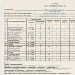
| Parameter name | Meaning |
| Article subject: | Protective function |
| Rubric (thematic category) | cooking |
Allows content to slide from top to bottom
BIBLIOGRAPHY
CONCLUSIONS
Τᴀᴋᴎᴍ ᴏϬᴩᴀᴈᴏᴍ, the main personal qualities of an entrepreneur are: independence; ambition; persistence; diligence; durability. The presence of such personality traits is one of the essential conditions success.
In addition to the actual personal qualities, an entrepreneur must have a set of specific knowledge, skills and abilities in the area in which he works. It is clear that in order to successfully financial transactions an entrepreneur needs at least minimum set knowledge in the financial and credit area and accounting͵ and a person who decides to organize the production of furniture must have a minimum technical education. However, these limitations are not definitive. It often happened that an entrepreneur received special knowledge and skills already in the course of developing his business, and at its first stages he acted either intuitively or with the help of attracted specialists. The main thing here is the desire to learn and improve one's skills in order to improve one's business, and such a desire already applies to personal qualities(curiosity, perseverance, ambition).
The study of the personality of an entrepreneur with the help of psychological tests not only helps to clarify certain aspects of his personality, but also shows in which direction he should work on himself in order to increase the efficiency of his entrepreneurial activity.
Akperov I. G., Maslikova Zh. V. Psychology of entrepreneurship. - M: Finance and statistics, 2003.
Zavyalova E.K., Posokhova S.T. Psychology of Entrepreneurship: Tutorial. - St. Petersburg: Ed. St. Petersburg State University, 2004.
Meneghetti A. Psychology of a leader. - M., 2001. - S. 15.
Platonov K.K. Structure and development of personality. - M.: Nauka, 1986. S. 24.
Entrepreneurship: Textbook / Ed. M. L. Lapusty. - M.: INFRA-M, 2003.
Steven J. Train Your Dragons. - St. Petersburg: Peter-press, 1996.
Shcherbatykh Yu.V. Psychology of Entrepreneurship and Business: Textbook. - St. Petersburg: Peter, 2008. S. 45.
Shcherbatykh Yu. V. Psychology of success. - M.: Eksmo, 2005.
The mucosa is fairly smooth
Lubricated with mucus (produced by the mucous glands of the shell itself)
Mucus - envelops m / o, viscosity does not allow penetration into the bloodstream
Accumulation of lymphoid tissue - consists of lymphocytes of varying degrees of maturity. Lymphoid tissue forms clusters:
ü Tonsils - located at the very beginning of the digestive and respiratory tubes:
o Palatine tonsils - on both sides of the pharynx
o Lingual - in the region of the root of the tongue
o Pharyngeal tonsil - m / at the upper and rear wall of the nasopharynx (vault) under the tuberculum faringeum
o Tubal tonsils - near the pharyngeal opening of the auditory tube
ü Single follicles - located throughout the bp, their total weight is about 2 kg;
ü Lymphoid plaques - contain dozens of lymphocytes, are present only in the ileum - Peyer's patches, their number is about 20-30
ü Vermiform appendix - its mucosa contains lymphoid tissue. it intestinal tonsil.
· Alternation of different media throughout the alimentary canal.
With the weakening of protective devices, immunity decreases !!!
- chemical processing of food- carried out by digestive juices, which are produced by the digestive glands. Throughout the p.t. there are glands:
By size:
Large
Major salivary glands (parotid, submandibular, sublingual)
Liver - produces bile that enters the duodenum
Pancreas - pancreatic juice, insulin.
Minor salivary glands (labial, buccal, palatine, lingual)
Gastric glands
Intestinal glands - in the lining of the small intestine
By localization:
In the thickness of the mucous membrane
Small salivary
Gastric
Glands of the jejunum and ileum of the small intestine
under the mucous layer
Duodenal glands
Outside the digestive tube
All large glands
Chemical processing in oral cavity- saliva, in the stomach - gastric juice, 12pc - bile, pancreatic juice. and glands of the 12pc itself, in the jejunum and ileum - under the influence of their own juices. Chemical processing ends in the small intestine. In the colon, fiber is broken down under the influence of microorganisms (m / o).
- absorption of nutrients – nutrients absorbed into the blood and lymphatic vessels. Absorption begins:
In the oral cavity (dr. Wed, alcohol)
Stomach (l / s, alcohol, nutrients)
The small intestine is the main absorption process
Large intestine - mostly water is absorbed
The small intestine is long, its mucosa has:
1. Circular folds, they increase the suction surface. On the border between the departments form valves
2. Villi - from 1.5 to 4 million, height 1mm, the wall is very thin.
3. Crypts - deepening of the mucosa
4. Epithelial cells have outgrowths - microvilli (up to 300 per cell).
Τᴀᴋᴎᴍ ᴏϬᴩᴀᴈᴏᴍ, mucosal area 1500 m2.
submucosal layer. Consists of loose connective tissue. Purpose:
Fixes the mucous membrane to the muscle;
Provides mobile fixation - the mucous membrane forms folds
Vessels and nerves pass
Muscular sheath. Formed by smooth muscle tissue. But around the oral cavity, the muscles of the pharynx, the upper third of the esophagus, the lower part of the rectum are striated.
The muscular layer of the digestive tube forms two layers:
Longitudinal - external)
shortens the alimentary canal
Straightens curves
Transverse (circular) - internal
Provides peristalsis - wavy narrowing of the intestinal lumen
Forms sphincters - local thickenings between the departments of the p.t. (esophagus - stomach, stomach - 12 pcs, small intestine - large intestine, in the lower part of the rectum).
Sphincters are strengthened by valves - against the sphincter, the mucous membrane forms a circular fold. In the mucous membrane under the valves there are venous plexuses.
Sphincter + Valve + Venous plexus = closing apparatus.
Purpose: prevention of premature emptying of the outgoing department; prevents content from being pushed back.
Only the stomach has three layers (+ oblique layer), as it acts as a reservoir and mixes food. Three layers also have a uterus, bladder, heart - the reservoir must be completely emptied.
Outer shell.
Connective tissue membrane - not in the abdominal cavity: pharynx, esophagus, rectum outside. Consists of a loose connective tissue sheath:
Fixes organs to bones
Connects organs to each other. There are no voids between organs, it is filled with loose connective tissue
Provides organ mobility - provides functional organ mobility
Vessels and nerves pass through it (in the adventitial layers)
The serous membrane is the organs of the abdominal cavity, formed by the peritoneum. The same purpose as the connection-woven sheath.
Protective function - concept and types. Classification and features of the category "Protective function" 2017, 2018.
Protective proteins
Protective proteins allow you to protect the body from the invasion of attacking bacteria, viruses and from the penetration of foreign proteins (the generalized name of foreign bodies is antigens).
The role of protective proteins is performed by immunoglobulins (their other name is antibodies), they recognize antigens that have penetrated the body and firmly bind to them.
In the body of mammals, including humans, there are five classes of immunoglobulins: M, G, A, D and E, their structure, as the name implies, is globular, in addition, they are all built in a similar way. The molecular organization of antibodies is shown on the slide using class G immunoglobulin as an example. The molecule contains four polypeptide chains joined by three disulfide bridges S-S(they are shown on the slide) with thickened valence bonds and large characters S ), in addition, each polymer chain contains intrachain disulfide bridges .
Two large polymer chains (highlighted in blue) contain 400–600 amino acid residues.
The other two chains (highlighted in green) are almost half as long, containing approximately 220 amino acid residues. All four chains are located in such a way that the terminal H 2 N-groups are directed in one direction.
After the body comes into contact with a foreign protein (antigen), the cells of the immune system begin to produce immunoglobulins (antibodies), which accumulate in the blood serum. At the first stage, the main work is done by chain sections containing terminal H 2 N (in Fig. 27, the corresponding sections are marked in light blue and light green). These are antigen capture sites. In the process of immunoglobulin synthesis, these sites are formed in such a way that their structure and configuration correspond as much as possible to the structure of the approaching antigen (like a key to a lock, like enzymes, but the tasks in this case are different). Thus, for each antigen, a strictly individual antibody is created as an immune response. Not a single known protein can change its structure so “plastically” depending on external factors, in addition to immunoglobulins. Enzymes solve the problem of structural conformity to the reagent in a different way - with the help of a gigantic set of various enzymes for all possible cases, and immunoglobulins each time rebuild the "working tool". Moreover, the hinge region of the immunoglobulin provides the two capture regions with some independent mobility, as a result, the immunoglobulin molecule can immediately “find” the two most convenient regions for capture in the antigen in order to securely fix it, this resembles the actions of a crustacean creature.
Next, a chain of successive reactions of the body's immune system is turned on, immunoglobulins of other classes are connected, as a result, the foreign protein is deactivated, and then the antigen (foreign microorganism or toxin) is destroyed and removed.
After contact with the antigen, the maximum concentration of immunoglobulin is reached (depending on the nature of the antigen and the individual characteristics of the organism itself) within a few hours (sometimes several days). The body retains the memory of such contact, and when attacked again with the same antigen, immunoglobulins accumulate in the blood serum much faster and in greater quantities - acquired immunity occurs.
The above classification of proteins is to a certain extent conditional, for example, the thrombin protein, mentioned among protective proteins, is essentially an enzyme that catalyzes the hydrolysis of peptide bonds, that is, it belongs to the class of proteases.
to protective proteins often referred to as proteins snake venom and toxic proteins some plants because their task is to protect the body from damage.
There are proteins whose functions are so unique that it makes it difficult to classify them. For example, the protein monellin, found in an African plant, is very sweet tasting and has been the subject of study as a non-toxic substance that can be used in place of sugar to prevent obesity. The blood plasma of some Antarctic fish contains proteins with antifreeze properties that keep the blood of these fish from freezing.
Proteins of the blood coagulation system have protective properties e.g. fibrinogen, thrombin. They are involved in the formation of a blood clot, which clogs the damaged vessel and prevents blood loss.
5 Contractile and motor proteins give the body the ability to contract, change shape and move, primarily, we are talking about muscles. 40% of the mass of all proteins contained in the muscles is myosin (mys, myos, Greek. - muscle). Its molecule contains both fibrillar and globular parts.
Such molecules combine into large aggregates containing 300–400 molecules.
When the concentration of calcium ions changes in the space surrounding the muscle fibers, a reversible change in the conformation of the molecules occurs - a change in the shape of the chain due to the rotation of individual fragments around the valence bonds. This leads to muscle contraction and relaxation, the signal to change the concentration of calcium ions comes from the nerve endings in the muscle fibers. Artificial muscle contraction can be caused by the action of electrical impulses, leading to a sharp change in the concentration of calcium ions, this is the basis for stimulating the heart muscle to restore the work of the heart.
Due to sliding relative to each other actin ( actins) and myosin ( myosins) protofibrils, muscle contraction occurs, as well as non-muscle intracellular contractions. The movement of cilia and flagella is associated with the sliding of microtubules relative to each other, which are of a protein nature.
Some Arctic and Antarctic fish contain proteins in the blood - antifreezes that prevent it from freezing.
Some proteins, when performing their functions, endow the cell with the ability to either contract or move. These proteins include actin and myosin, fibrillar proteins involved in skeletal muscle contraction. Another example of such proteins is tubulin, from which cell organelles, microtubules, are built. Microtubules regulate the separation of chromatids during cell division. Microtubules are important elements of cilia and flagella, with the help of which cells move.
However, there is a large number of proteins that have unique functions that are not included in this rather simple classification.
6 Regulatory proteins, more often called hormones, are involved in various physiological processes.
Regulatory proteins are large group protein hormones involved in maintaining the constancy of the internal environment of the body, which act on specific target cells.
Many hormones are oligopeptides or proteins (eg, insulin, glucagon [an insulin antagonist], adrenocorticotropic hormone, etc.).
The hormone insulin consists of two α-chains connected by disulfide bridges.
Insulin is a hormone produced in the cells of the islets of Langerhans in the pancreas. It plays a critical role in the metabolism of glucose in the blood.
In addition, regulatory proteins include proteins whose attachment to other proteins or other cell structures regulates their function. For example, the calmodulin protein in complex with four Ca2+ ions can attach to some enzymes, changing their activity.
Regulatory DNA-binding proteins, attaching at certain moments to specific DNA regions, can regulate the rate of reading genetic information.
The pituitary gland of the brain synthesizes a hormone that regulates the growth of the body. There are regulatory proteins that control the biosynthesis of various enzymes in the body.
The figure shows - INSULIN PROTEIN - in the form of a three-dimensional model and in the form of a tertiary structure. Consists of two α-helical chains connected by two disulfide bridges (compare with Fig. 2, where its structure is shown schematically)
INSULIN MOLECULE, built from 51 amino acid residues, fragments of the same amino acids are marked with the corresponding background color. The cysteine amino acid residues (abbreviated designation CIS) contained in the chain form disulfide bridges -S-S-, which link two polymer molecules, or form jumpers within one chain.
Receptor ( signal) protein function
Some proteins are embedded in cell membrane, are able to change their structure under the influence of the external environment.
This is how signals are received from the outside and information is transmitted to the cell.
An example would be phytochrome- a photosensitive protein that regulates the photoperiodic response of plants, and opsin - component rhodopsin pigment - , an integral membrane protein found in the cells of the retina.
Phytochrome (from Phyto... and Greek chroma - color, paint) blue pigment from the group of complex proteins - chromoproteins; present in the cells of photosynthetic organisms. It was first discovered by the American biochemist W. Butler in 1959 in the cotyledons of turnip seedlings grown in the dark.
Bluish phytochromes are photosynthetically inactive pigments.
However, it has been established that the synthesis of biopolymers (DNA, RNA, proteins), systems for the biosynthesis of chlorophyll, carotenoids, anthocyanins, organic phosphates, and vitamins are under the control of phytochrome. F. accelerates the catabolic breakdown of polysaccharides, fats, and reserve proteins, and activates cellular respiration and oxidative phosphorylation.
The enzymes exist in two interconvertible forms, F660 and F730, which differ in their absorption spectra. Under the action of red light with a wavelength of λ = 660 nm, inactive Ф660 is converted into active Ф730. The reverse transformation occurs either in the dark or when illuminated with red light with λ = 730 nm. It is believed that these interconversions are due to cis-trans isomerization of the F chromophore and conformational rearrangements of the protein.
Signal molecules (hormones, neurotransmitters) act on intracellular processes through interaction with specific receptor proteins.
Hormones circulating in the blood find target cells and act on them by specifically binding to receptor proteins, usually embedded in the cell membrane. For hydrophobic regulatory molecules passing through the cell membrane, the receptors are localized in the cytoplasm of the cells.
Signal molecules (hormones, neurotransmitters) act on intracellular processes through interaction with specific receptor proteins. Thus, hormones circulating in the blood find target cells and act on them by specifically binding to receptor proteins, usually embedded in the cell membrane. For hydrophobic regulatory molecules passing through the cell membrane, the receptors are localized in the cytoplasm of the cells.
The most important of these are phytochromes A and B (phyA and phyB). Phytochrome A
Performs many different photoregulatory functions. With its participation, stimulation and inhibition of seed germination, induction of de-etiolation, regulation of the synthesis of various enzymes, regulation of root development, stimulation of flowering and regulation of circadian rhythms occur.


Cycle of major changes in rhodopsin in retinal rods
RHODOPSIN (from the Greek rhodon - rose and opsis - vision), visual purple, osn. visual pigment of retinal rods of vertebrates (except for some fish and amphibians on early stages development) and invertebrates.
According to chem. In nature, rhodopsin is a complex protein (chromoprotein), which includes 11-cis-retinal (chromophore group), a glycoprotein, that is, a protein combined with sugars, and lipids (the so-called opsin part). Mol. the mass of rhodopsin of vertebrates is approx. 40,000, cephalopods - approx. 70 000. R. - main. structural and functional component of the outer segment of the rods (see Vision, Retina, Photoreceptors).
The visual act begins with R.'s absorption of a quantum of light (the maximum absorption spectrum of R. is approx. 500 nm). At the same time, 11-cis-retinal isomerization occurs in a completely trans-form (see formulas), which leads to a gradual decomposition (photolysis) of the R. molecule, a change in ion transport in the photoreceptor and the appearance of an electric. signal, to-ry is transmitted to the nerve elements of the retina. Regeneration of R. is carried out either by synthesis from 11-cis-retinal and opsin released after photolysis, or by absorption of the second quantum by one of the intermediate products of photolysis, as well as in the process of synthesis of new disks of the outer segment of the retina ( last way for sticks basic).
In the cell membranes of some halophilic bacteria, a pigment was found, which also includes retinal, glycoprotein, and lipids. This bacterial radapsin (its structure has not been fully established) apparently participates in photosynthesis along with other bacterial pigments.
Of particular importance for the action of phytochrome is its reversibility: this chromoprotein (a complex protein containing, in addition to amino acids, also coloring components) occurs in two forms that can be converted one into another.
Blue phytochrome 660 (Ф 660) has an absorption maximum in the light red region of the spectrum with a wavelength of 660 nm, and green-blue phytochrome 730 (Ф 730) - in the dark red region of the spectrum with a wavelength of 730 nm. 
When illuminated with light red light, inactive F 660 is converted into physiologically active F 730, and when illuminated with dark red light, F 730 is converted into F 660.
8 Dietary and storage proteins, as the name implies, serve as sources of internal nutrition, more often for the embryos of plants and animals, as well as in the early stages of development of young organisms.
Dietary proteins are albumen- the main component of egg white, as well as casein is the main protein in milk.
Under the action of an enzyme pepsin casein curdles in the stomach, which ensures its retention in the digestive tract and efficient absorption. Casein contains fragments of all the amino acids needed by the body.
In ferritin, which is found in animal tissues, iron ions are stored.
Storage proteins are also myoglobin, similar in composition and structure to hemoglobin. myoglobin focused mainly on in the muscles, its main role is oxygen storage, which gives him hemoglobin. It quickly saturates with oxygen (much faster than hemoglobin), and then gradually transfers it to various tissues during subsequent exercise and oxygen deficiency free him..
All this variety of functions stems from a very simple set of 20 amino acids, from which the polypeptide chain of a protein is built. It is the different amounts and different combinations of these amino acids in the chain that determine the uniqueness of a particular protein.
Like other biological macromolecules (polysaccharides, lipids and nucleic acids), proteins are essential components of all living organisms and play a decisive role in cell life. Proteins carry out metabolic processes. They are part of intracellular structures - organelles and cytoskeleton, secreted into the extracellular space, where they can act as a signal transmitted between cells, participate in the hydrolysis of food and the formation of intercellular substance.
The classification of proteins according to their functions is rather arbitrary, since the same protein can perform several functions. A well-studied example of such multifunctionality is lysyl-tRNA synthetase, an enzyme from the class of aminoacyl-tRNA synthetases, which not only attaches a lysine residue to tRNA, but also regulates the transcription of several genes. Proteins perform many functions due to their enzymatic activity. So, enzymes are the motor protein myosin, the regulatory proteins of protein kinase, the transport protein sodium-potassium adenosine triphosphatase, etc.
Molecular model of the urease enzyme of a bacterium Helicobacter pylori
catalytic function
Most well known function proteins in the body - catalysis of various chemical reactions. Enzymes are proteins that have specific catalytic properties, that is, each enzyme catalyzes one or more similar reactions. Enzymes catalyze reactions that break down complex molecules (catabolism) and synthesize them (anabolism), including DNA replication and repair and RNA template synthesis. By 2013, more than 5,000 enzymes had been described. The acceleration of the reaction as a result of enzymatic catalysis can be enormous: for example, the reaction catalyzed by the enzyme orotidine-5 "-phosphate decarboxylase proceeds 10 17 times faster than the non-catalyzed one (the half-reaction period for the decarboxylation of orotic acid is 78 million years without the enzyme and 18 milliseconds with the participation of the enzyme) Molecules that attach to an enzyme and change as a result of the reaction are called substrates.
Despite the fact that enzymes usually consist of hundreds of amino acid residues, only a small part of them interact with the substrate, and even fewer - on average 3-4 amino acid residues, often located far apart in the primary structure - are directly involved in catalysis. . The part of the enzyme molecule that provides substrate binding and catalysis is called the active site.
The International Union of Biochemistry and Molecular Biology in 1992 proposed the final version of the hierarchical nomenclature of enzymes based on the type of reactions they catalyze. According to this nomenclature, the names of enzymes must always have the ending - aza and be formed from the names of catalyzed reactions and their substrates. Each enzyme is assigned an individual code, by which it is easy to determine its position in the hierarchy of enzymes. According to the type of catalyzed reactions, all enzymes are divided into 6 classes:
- EC 1: Oxidoreductases catalyzing redox reactions;
- EC 2: Transferases that catalyze the transfer of chemical groups from one substrate molecule to another;
- EC 3: Hydrolases catalyzing the hydrolysis of chemical bonds;
- EC 4: Lyases that catalyze the breaking of chemical bonds without hydrolysis with the formation of a double bond in one of the products;
- EC 5: Isomerases that catalyze structural or geometric changes in the substrate molecule;
- EC 6: Ligases that catalyze the formation of chemical bonds between substrates by hydrolysis of the diphosphate bond of ATP or a similar triphosphate.
structural function
More: Structural function of proteins, fibrillar proteins
Structural proteins of the cytoskeleton, like a kind of armature, give shape to cells and many organelles and are involved in changing the shape of cells. Most structural proteins are filamentous: actin and tubulin monomers, for example, are globular, soluble proteins, but after polymerization they form long filaments that make up the cytoskeleton, enabling the cell to maintain its shape. Collagen and elastin are the main components of the intercellular substance of connective tissue (for example, cartilage), and hair, nails, bird feathers, and some shells are made up of another structural protein, keratin.

Protective function
More: Protective function of proteins
There are several types of protective functions of proteins:
- Physical protection. Physical protection of the body is provided by Collagen - a protein that forms the basis of the intercellular substance of connective tissues (including bones, cartilage, tendons and deep layers of the skin (dermis)); keratin, which forms the basis of horny shields, hair, feathers, horns, and other derivatives of the epidermis. Usually such proteins are considered as proteins with structural function. Examples of proteins of this group are fibrinogens and thrombins involved in blood coagulation.
- Chemical protection. The binding of toxins to protein molecules can provide their detoxification. A particularly decisive role in detoxification in humans is played by liver enzymes that break down poisons or convert them into a soluble form, which contributes to their rapid removal from the body.
- Immune protection. Proteins that make up blood and other biological fluids are involved in the body's defense response to both damage and attack by pathogens. Proteins of the complement system and antibodies (immunoglobulins) belong to the proteins of the second group; they neutralize bacteria, viruses or foreign proteins. Antibodies that are part of the adaptive immune system attach to foreign ones for given organism substances, antigens, and thereby neutralize them, directing them to the places of destruction. Antibodies can be secreted into the intercellular space or become attached to the membranes of specialized B-lymphocytes called plasma cells.
Regulatory function
More: Activator (proteins), Proteasome, Regulatory function of proteins
Many processes inside cells are regulated by protein molecules, which serve neither as a source of energy nor building material for the cell. These proteins regulate cell progression through the cell cycle, transcription, translation, splicing, the activity of other proteins, and many other processes. The regulatory function of proteins is carried out either due to enzymatic activity (for example, protein kinases), or due to specific binding to other molecules. Thus, transcription factors, activator proteins and repressor proteins, can regulate the intensity of gene transcription by binding to their regulatory sequences. At the level of translation, the reading of many mRNAs is also regulated by the addition of protein factors.
The most important role in the regulation of intracellular processes is played by protein kinases and protein phosphatases - enzymes that activate or inhibit the activity of other proteins by attaching to them or removing phosphate groups.

Signal function
More: Protein signaling function, Hormones, Cytokines
The signaling function of proteins is the ability of proteins to serve as signaling substances, transmitting signals between cells, tissues, organs and organisms. The signaling function is often combined with the regulatory function, since many intracellular regulatory proteins also carry out signal transduction.
The signal function is performed by proteins-hormones, cytokines, growth factors, etc.
Hormones are carried in the blood. Most animal hormones are proteins or peptides. The binding of a hormone to its receptor is a signal that triggers a cell response. Hormones regulate the concentration of substances in the blood and cells, growth, reproduction and other processes. An example of such proteins is insulin, which regulates the concentration of glucose in the blood.
Cells interact with each other using signal proteins transmitted through the intercellular substance. Such proteins include, for example, cytokines and growth factors.
Cytokines are peptide signaling molecules. They regulate interactions between cells, determine their survival, stimulate or suppress growth, differentiation, functional activity and apoptosis, ensure the coordination of actions of the immune, endocrine and nervous systems. An example of cytokines is tumor necrosis factor, which transmits inflammatory signals between body cells.
transport function
More: Transport function of proteins

Soluble proteins involved in the transport of small molecules must have a high affinity (affinity) for the substrate when it is present in high concentration, and it is easy to release it in places of low substrate concentration. An example of transport proteins is hemoglobin, which carries oxygen from the lungs to other tissues and carbon dioxide from tissues to lungs, and besides this, homologous proteins found in all kingdoms of living organisms.
Some membrane proteins are involved in the transport of small molecules through the cell membrane, changing its permeability. The lipid component of the membrane is waterproof (hydrophobic), which prevents the diffusion of polar or charged (ions) molecules. Membrane transport proteins are commonly classified into channel proteins and carrier proteins. Channel proteins contain internal water-filled pores that allow ions (via ion channels) or water molecules (via aquaporins) to move across the membrane. Many ion channels are specialized for the transport of only one ion; thus, potassium and sodium channels often distinguish between these similar ions and allow only one of them to pass through. Carrier proteins bind, like enzymes, every molecule or ion they carry and, unlike channels, can actively transport using the energy of ATP. The "powerhouse of the cell" - ATP synthase, which carries out the synthesis of ATP due to the proton gradient, can also be attributed to membrane transport proteins.
Spare (backup) function
These proteins include the so-called reserve proteins, which are stored as a source of energy and substances in plant seeds (for example, 7S and 11S globulins) and animal eggs. A number of other proteins are used in the body as a source of amino acids, which in turn are precursors of biologically active substances that regulate metabolic processes.
![]()
Receptor function
More: Cell receptor
Protein receptors can be located both in the cytoplasm and embedded in the cell membrane. One part of the receptor molecule receives the signal, which is often Chemical substance, and in some cases - light, mechanical action (for example, stretching) and other stimuli. When a signal is applied to a certain part of the molecule - the receptor protein - its conformational changes occur. As a result, the conformation of another part of the molecule, which transmits the signal to other cellular components, changes. There are several signaling mechanisms. Some receptors catalyze a specific chemical reaction; others serve as ion channels that open or close when a signal is applied; still others specifically bind intracellular messenger molecules. In membrane receptors, the part of the molecule that binds to the signal molecule is located on the cell surface, while the signal-transmitting domain is inside.
Motor (motor) function
A whole class of motor proteins provides body movements, for example, muscle contraction, including locomotion (myosin), movement of cells within the body (for example, amoeboid movement of leukocytes), movement of cilia and flagella, and in addition, active and directed intracellular transport (kinesin, dynein ). Dyneins and kinesins transport molecules along microtubules using ATP hydrolysis as an energy source. Dyneins carry molecules and organelles from the peripheral parts of the cell towards the centrosome, kinesins - in the opposite direction. Dyneins are also responsible for the movement of cilia and flagella in eukaryotes. Cytoplasmic variants of myosin can take part in the transport of molecules and organelles through microfilaments.
The main and, in a sense, unique biological functions of proteins that are uncharacteristic or only partially inherent in other classes of biopolymers include the following functions.
Structural (support) function
Collagen fibers perform a supporting function. (electron microscopy)
Proteins that do structural function, predominate among other proteins of the human body. Fibrillar proteins form the substance of the connective tissue - collagen, elastin (in the vascular wall of elastic-type vessels), keratin (in the skin and its derivatives), proteoglycans.
Enzymatic (catalytic) function
All enzymes are proteins that determine the rate of chemical reactions in biological systems. But at the same time, there are experimental data on the existence of ribozymes, that is, ribonucleic acid with catalytic activity, and abzymes - both mono- and polyclonal antibodies.
Receptor and hormonal function
transport function
Only proteins carry out the transport of substances in the blood, for example, lipoproteins (fat transport), hemoglobin (oxygen transport), transferrin (iron transport). Proteins transport calcium, magnesium, iron, copper and other ions in the blood.
The transport of substances through membranes is carried out by proteins - Na +, K + -ATPase (anti-directional transmembrane transfer of sodium and potassium ions), Ca 2+ -ATPase (pumping calcium ions out of the cell), glucose transporters.
Reserve (nutritional) function
This function is performed by the so-called reserve proteins. An example of a deposited protein is the production and accumulation of egg albumin (ovalbumin) in the egg. Animals and humans do not have such specialized depots, but during prolonged starvation, proteins of muscles, lymphoid organs, epithelial tissues and liver are used. The main protein of milk (casein) also performs a mainly nutritional function.
contractile function
There are a number of intracellular proteins designed to change the shape of the cell and the movement of the cell itself or its organelles. main role actin and myosin, specific proteins of muscle tissue, play in the processes of movement, and tubulin, a protein of the cytoskeleton, which provides the finest processes of cell vital activity - the divergence of chromosomes during mitosis.
Protective function
Functions of blood proteins
In the regulation of plasma protein content at a certain level great importance has a liver that fully synthesizes fibrinogen and blood albumins, mostα- and β-globulins, cells of the reticuloendothelial system of the bone marrow and lymph nodes.